Tuesday, November 30, 2004


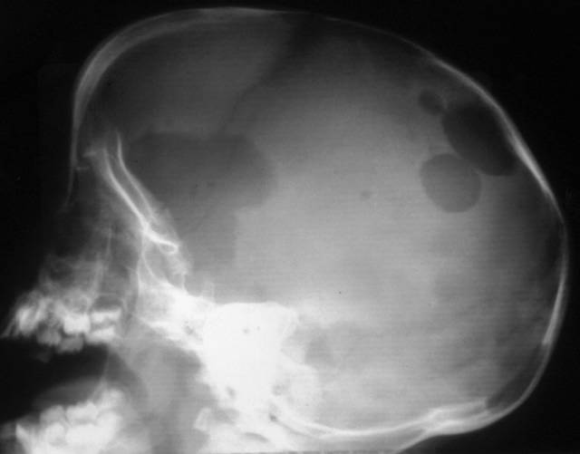
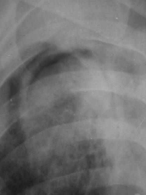
The "classical air-crescent sign"
Diagnosis- fungal ball or Aspergilloma!!!
COPYRIGHT (SUMER)

Saturday, November 27, 2004


classic appearance of a neurogenic bladder- known as "pine-tree appearance" also note the left Vesicoureteric reflux!!!
COPYRIGHT (SUMER)

Wednesday, November 24, 2004

journal watch

High resolution volume imaging of airways and lung parenchyma with multislice CT.

Chooi WK, Morcos SK.Department of Diagnostic Imaging, Northern General Hospital, Sheffield Teaching Hospitals NHS Trust, Herries Road, Sheffield S5 7AU, UK.


The value of multislice CT (MSCT) in imaging the peripheral airways and lung parenchyma has not been widely investigated. In this article the authors' experience in the use of MSCT (4-slice scanner) in imaging patients with suspected parenchymal lung disease or airways abnormalities will be presented. The technique described should be modified with the more modern 8-slice or 16-slice scanners. The whole thorax is scanned contiguously using 4 x 1 mm collimation from the lung bases up to apices in end-inspiration while the patient is in the prone position. Collimation of 2 x 0.5 mm is used at 8-10 levels evenly spaced in expiratory scans and also in the breathless patient who is scanned during gentle breathing. High resolution images of the lungs (1 mm slice thickness) are reconstructed in the following planes: axial (10 mm apart from apices to bases), coronal (six evenly spaced through the chest) and sagittal (four images evenly spaced through each lung). Paddlewheel reconstruction is used if further assessment of the airways is required, and three-dimensional imaging is used mainly for assessment of the trachea and major bronchi. Contiguous axial images (10 mm slice thickness) of the whole lung and mediastinum are also produced and referred to as a screenogram. Axial images of 1 mm slice thickness are produced with expiratory scans and for breathless patients. All the images are produced independently by the radiographic staff and are provided as hard copies (20 frames/film) for reporting. However, if facilities are adequate, direct reporting from the workstation is more effective in reviewing large number of images. The technique is effective in assessment of infiltrative lung disease, emphysema, bronchiectasis and central airways. The screenogram offers comprehensive evaluation of the lung and mediastinum, but the radiation dose associated with high resolution volume imaging remains a source of concern.


Br J Radiol. 2004 Dec;77 Suppl 1:S98-S105.


radiology news

MR detects pancreatic islet inflammation in type 1 diabetes


CONTEXT: In type 1 diabetes, the body's immune system mistakenly launches an attack on insulin-producing beta cells, sending T cells to invade pancreatic islets. Until recently, physicians could track type 1 diabetes only by monitoring blood levels of antibodies directed against pancreatic islet proteins. The test, however, usually detected type 1 diabetes late in its progression, after most islet beta cells had been destroyed and autoimmune processes had been played out. Researchers at Massachusetts General Hospital and Boston's Joslin Diabetes Center are using magnetofluorescence contrast nanoparticles, monitored with high-field MR, to devise a better test. It measures the permeability of the small blood vessels surrounding and within the islets, an early marker of this inflammation.


RESULTS: The technique has been successfully tested on a mouse model. For imaging, long-circulating magnetofluorescent nanoparticles were used. They consist of a superparamagnetic iron oxide (SPIO) core, a crosslinked dextran coating, and amino groups to which Alexa-488 fluorochrome is attached. Transgenic mice were imaged with an 8.5T micro-MR scanner 24 hours after contrast injection. Contrast accumulation was monitored in vivo, and a positive correlation between probe accumulation and insulitis was documented.


IMAGE: Higher MR relativities were measured in the pancreas of normal mice compared with diabetic mice 24 hours after contrast injection. (Image reprinted with permission of Proceedings of the National Academy of Science)


IMPLICATIONS: This imaging strategy may prove invaluable in helping identify early insulitis and for monitoring therapeutic interventions aimed at stopping its progression. MGH has already safely used the method in human clinical trials to detect the spread of prostate cancers to the lymph nodes.


u get the latest on sumer's radiology site


Saturday, November 20, 2004

AIIMS NOV 2004 FULLY SOLVED!!!


aiims nov 2004

cost-150/-

new concept-online support with mcq discussion board

www.sumerdoc.blogspot.com


highlights-extensive flowcharts and diagrams with special emphasis on protocols. beautiful explaination of the flow-volume curve with to the point diagrams.


features-fully solved and explained by sumer sethi and sidharth sethi is available in the market...highlights of the book are authentic references including harrisons 16th edition, diagrams and flowcharts, extensive coverage of topics so that all repeat questions are tackled...and first book of its kind to give extensive online support with an online discussion forum support by the author.. also by the same author "review of radiology" solving quite a few questions in this aiims!! and may 2004 aiims fully solved peepee publishers..one of the main features of this book is in the reference the author has not modified the exact language of the text taken from standard textbooks so that it is very reliable not biased by personal beliefs.. personal beliefs or fundas built without text basis are usually disastrous for mcq exams so emphasis has been kept on what the latest and most standard books say.. wherever possible and required multiple references have been given...

COPYRIGHT (SUMER)


Acute appendicitis. Ultrasound examination of the right lower quadrant demonstrates a tubular, hyporeflective, noncompressible structure with diameter of 8 mm. Hyporeflective aspect of adjacent fatty tissue is due to inflammatory oedema.

COPYRIGHT (SUMER)

Friday, November 19, 2004

journal watch

Pulmonary Nodules on Multi-Detector Row CT Scans: Performance Comparison of Radiologists and Computer-aided Detection.

Rubin GD, Lyo JK, Paik DS, Sherbondy AJ, Chow LC, Leung AN, Mindelzun R, Schraedley-Desmond PK, Zinck SE, Naidich DP, Napel S.Departments of Radiology and Electrical Engineering, Stanford University School of Medicine, 300 Pasteur Dr, S-072, Stanford, CA 94305-5105.

PURPOSE: To compare the performance of radiologists and of a computer-aided detection (CAD) algorithm for pulmonary nodule detection on thin-section thoracic computed tomographic (CT) scans.

MATERIALS AND METHODS: The study was approved by the institutional review board. The requirement of informed consent was waived. Twenty outpatients (age range, 15-91 years; mean, 64 years) were examined with chest CT (multi-detector row scanner, four detector rows, 1.25-mm section thickness, and 0.6-mm interval) for pulmonary nodules. Three radiologists independently analyzed CT scans, recorded the locus of each nodule candidate, and assigned each a confidence score. A CAD algorithm with parameters chosen by using cross validation was applied to the 20 scans. The reference standard was established by two experienced thoracic radiologists in consensus, with blind review of all nodule candidates and free search for additional nodules at a dedicated workstation for three-dimensional image analysis. True-positive (TP) and false-positive (FP) results and confidence levels were used to generate free-response receiver operating characteristic (ROC) plots. Double-reading performance was determined on the basis of TP detections by either reader.

RESULTS: The 20 scans showed 195 noncalcified nodules with a diameter of 3 mm or more (reference reading). Area under the alternative free-response ROC curve was 0.54, 0.48, 0.55, and 0.36 for CAD and readers 1-3, respectively. Differences between reader 3 and CAD and between readers 2 and 3 were significant (P < .05); those between CAD and readers 1 and 2 were not significant. Mean sensitivity for individual readings was 50% (range, 41%-60%); double reading resulted in increase to 63% (range, 56%-67%). With CAD used at a threshold allowing only three FP detections per CT scan, mean sensitivity was increased to 76% (range, 73%-78%). CAD complemented individual readers by detecting additional nodules more effectively than did a second reader; CAD-reader weighted kappa values were significantly lower than reader-reader weighted kappa values (Wilcoxon rank sum test, P < .05).

CONCLUSION: With CAD used at a level allowing only three FP detections per CT scan, sensitivity was substantially higher than with conventional double reading.


Radiology. 2004 Nov 10; [Epub ahead of print]

this is scary!!!!

Tuberculosis risk high among Indian resident physicians


Resident doctors in India have nearly nine times the risk of contracting tuberculosis from their patients than the general population has of contracting the disease, researchers from Chandigarh, India, report.

"Due to the exceptionally high burden of tuberculosis in the general population in India, it is expected that doctors caring for such patients have a high probability of acquiring the disease," Dr. KG Rao and colleagues write.

To assess this increased risk, Dr. KG Rao and colleagues from the Post Graduate Institute of Medical Education and Research evaluated 873 doctors in various stages of their residencies.

The resulting study population was divided into two groups: group one - comprised of 470 doctors who were already undergoing residency in January 2001 when the study began, and group two, comprising 231 residents who joined during 2001. Researchers administered a detailed questionnaire on their medical history, previous and present exposure to tuberculosis, and treatment to group one at the start of the study and to group two after completion one year of training.

Thirteen residents in both groups, including nine (1.9%) in group one and 4 (1.7%) in group two contacted tuberculosis during the course of residency, giving an overall risk of 17.3 per 1000, nearly nine times higher than the population risk in India, Dr. Rao and colleagues report in the November issue of International Journal of Tuberculosis and Lung Diseases.

Extrapulmonary tuberculosis was predominant, with six (67%) residents in group 1 and three (75%) from group 2 developing this severe form of the disease, the researchers add. The incidence of extrapulmonary tuberculosis was significantly higher in the general population, probably because of repeated contact and prior exposure to tuberculosis, they suggest.

As compared to a previous 40-year cohort study on tuberculosis risk in US physicians, the risk of tuberculosis among Indian residents was eight times higher, the authors note. This could be due to the low tuberculosis prevalence in the US, they postulate.

"The most appropriate method of preventing such transmission is effective treatment of smear-positive pulmonary tuberculosis patients with standard four-drug anti-tuberculosis therapy, as most patients become non-infectious after 2 weeks of treatment if the organism is drug-sensitive," the authors conclude.


Int J Tuberc Lung Dis 2004; 8:1392-1394.

Wednesday, November 17, 2004


MR, axial T2-weighted image, shows the absent vermis and hypoplasia of the cerebellar hemispheres and a large posterior fossa cyst!!
THE DANDY WALKER MALFORMATION!!!
COPYRIGHT (SUMER)

Tuesday, November 16, 2004

Sunday, November 14, 2004


MRCP image annular pancreas showing typical ductal loop!!!
COPYRIGHT (SUMER)

Saturday, November 13, 2004


biconvex extradural hematoma!!!
COPYRIGHT (SUMER)

journal watch

Is 'virtual histology' the next step after the 'virtual autopsy'? Magnetic resonance microscopy in forensic medicine.


AIM: The study aimed to validate magnetic resonance microscopy (MRM) studies of forensic tissue specimens (skin samples with electric injury patterns) against the results from routine histology.

METHODS AND RESULTS: Computed tomography and magnetic resonance imaging are fast becoming important tools in clinical and forensic pathology. This study is the first forensic application of MRM to the analysis of electric injury patterns in human skin. Three-dimensional high-resolution MRM images of fixed skin specimens provided a complete 3D view of the damaged tissues at the site of an electric injury as well as in neighboring tissues, consistent with histologic findings. The image intensity of the dermal layer in T2-weighted MRM images was reduced in the central zone due to carbonization or coagulation necrosis and increased in the intermediate zone because of dermal edema. A subjacent blood vessel with an intravascular occlusion supports the hypothesis that current traveled through the vascular system before arcing to ground.

CONCLUSION: High-resolution imaging offers a noninvasive alternative to conventional histology in forensic wound analysis and can be used to perform 3D virtual histology.


Magn Reson Imaging. 2004 Oct;22(8):1131-8.


Friday, November 12, 2004

Thursday, November 11, 2004

Wednesday, November 10, 2004


herpes encephalitis!! note the bilateral temporal and frontal involvement
COPYRIGHT (SUMER)

Sunday, November 7, 2004


golden S sign-- right upper lobe collapse with central mass..
COPYRIGHT (SUMER)

Friday, November 5, 2004

Tc 99 DECAY SCHEME


Tc 99 most commonly used for radioisotope imaging
COPYRIGHT (SUMER)

Thursday, November 4, 2004

Journal watch

CT Appearance of Acute Appendagitis

OBJECTIVE: Our aim was to describe the spectrum of CT findings in patients with acute epiploic appendagitis and also to evaluate the changes seen with this condition.

MATERIALS AND METHODS: Fifty patients diagnosed with acute epiploic appendagitis seen on contrast-enhanced CT were included in this study. The CT scans of the epiploic appendagitis were evaluated for the presence of colon wall thickening, a focal fatty center, inflammatory changes, location in relationship to the colon, size, and presence or absence of central high density within the fat. In 10 patients, the initial findings were compared with findings of follow-up CT performed between 3 days-21 months after the first CT.

RESULTS: The most common part of colon involved by acute epiploic appendagitis was the sigmoid colon (31/50), and the most common position was anterior to the colonic lumen (41/50). All 50 patients with acute epiploic appendagitis had a central fatty core surrounded by inflammation. Colon wall thickening was present in only two, and a central high-density focus was noted only in 27 of 50 patients. In 86% (43/50) of patients, the fatty central core was between 1.5 and 3.5 cm in length. The changes seen on follow-up CT varied, including increased density with a decrease in the size of the fatty central core, no change, complete resolution of findings, and minimal residual density.

CONCLUSION: On CT, acute epiploic appendagitis has a predictable appearance in terms of location, size, and density. The most common finding on CT is a fat-density oval lesion with surrounding inflammation on the anterior aspect of the sigmoid colon. The changes on CT are not predictable in the 2-week to 6-month window.

AJR Am J Roentgenol. 2004 Nov;183(5):1303-7.

Wednesday, November 3, 2004


Pulmonary angiography showing a Rasmussen's aneurysm of the right lower lobe incidentally discovered in a patient with a previous history of cavitary tuberculosis of the same lobe.

COPYRIGHT (SUMER)

Tuesday, November 2, 2004


phantom tumour-collection of pleural fluid between lobes, i.e. within the major and minor fissures. The radiographic appearance depends on the shape and orientation of the fissure, the volume of fluid, its position within the fissure and the radiographic projection. Interlobar fluid is particularly common in heart failure.

COPYRIGHT (SUMER)

Scurvy
Rare before 6 months of age since the storage of vitamin C in neonate is generally adequate
• Wimberger sign: presence of a sclerotic rim around epiphysis
• White line of frankel:dense zone of provisional calci­fication at the growing metaphysis
• Trumerfeld zone:a lucent zone below white line due to lack of mineralisation
• Pelkan spur:as the area is prone to fractures manifesting at cortical margin
• Osteoporosis
• Subperiosteal haemorrhage.
COPYRIGHT (SUMER)

Tutorials

STRING OF BEADS


PULMONARY EDEMA


BILIARY ATRESIA


MITRAL STENOSIS


MRI IN BLADDER CANCER


RADIOBIOLOGY


COPD X-RAY FINDINGS


WHITAKER TEST


CHYLOTHORAX


PULMONARY INFECTIONS


RADIOSENSTIVITY OF TUMOURS


DYSPHAGIA


ERCP VS MRCP


RADIATION UNITS


SCURVY


Tc99 decay scheme


Monday, November 1, 2004


Langerhans' cell histiocytosis

Lateral radiograph of the skull demonstrating multiple lytic lesions of histiocytosis. The anterior lesion has a "geographical" pattern.


COPYRIGHT (SUMER)

imaging news

Portable CT goes to ICU patients


Portable CT scanners can now provide valuable abdominal diagnostic information for intensive care unit patients without forcing them to leave the unit, according to a study in the September issue of the American Journal of Roentgenology.

The first portable CT scanners were deployed at several large institutions in the late 1990s. Little is known, however, about their performance and use in addition to cranial imaging, which remains portable CT's main application. Unlike cranial CT, an abdominal scan requires moving patients from their beds onto the scanning table. But portable scans acquired at the bedside can reduce well-documented risks associated with transport of ICU patients to other hospital locations.

Dr. Michael M. Maher and colleagues at Massachusetts General Hospital retrospectively reviewed spiral portable abdominal CT scans from 107 patients obtained between June 1999 and December 2000. They compared portable CT's image quality and diagnostic value with that of available stationary CT scans.

Although image quality from portable abdominal CT did not match stationary CT, the researchers found they were able to obtain important diagnostic information without moving patients from the ICU.

Researchers obtained 122 portable and 41 stationary CT scans, with 47 and 15, respectively, enhanced by contrast. Intravenous contrast improved portable CT scan quality. Quality scores for portable CT scans, however, were consistently lower than those for stationary CT, both with and without contrast.

Findings on portable CT confirmed 33 conditions suspected before scanning. Portable scanning also detected evidence of infection in 18 patients and hemorrhage in 16, led to seven laparotomies and six percutaneous drainage procedures, and influenced a change in patient management in 33 cases. Surgery or autopsy results confirmed portable CT findings in 12 of 17 cases.

The spiral portable CT scanning protocol included two 285 to 355-mm-long volumes acquired with 5-mm slice thickness during 70 sec at 120 to 130 kVp, 30 to 40 mAs, and a 1 to 1.5-sec pitch. Patient condition and imaging indication determined IV contrast use. Stationary scanning was performed with single- and four-detector spiral scanners.

Interpretation of portable CT studies must proceed with caution, however. Some portable CT parameters, such as accuracy and negative predictive value, were unknown. While portable scanning is useful in many cases, patients who require valuable diagnostic information should be moved to more sophisticated imaging installations whenever possible, researchers said.


Can MRI replace DMSA in the detection of renal parenchymal defects in children with urinary tract infections?

Background: Renal parenchymal defects may be a consequence of urinary tract infections (UTI) in childhood. MRI is a non-radiation imaging modality compared with DMSA scanning. Objective: To compare DMSA with MRI for the detection of renal parenchymal defects in children presenting for radiological investigation after a first UTI. Materials and methods: Both DMSA and MRI were performed at the same appointment in 37 children (aged 4 months-13 years; mean 4.5 years) with a history of UTI. Both planar and SPECT DMSA were performed. MRI of the kidneys employed axial and coronal T1-, T2- and fat-saturated T1-weighted (T1-W) sequences. Some children had imaging after IV contrast medium. Results: The coronal fat-saturated T1-W sequence was the best sequence and it detected all the findings on MRI. MRI had a sensitivity of 77% and a specificity of 87% for the detection of a scarred kidney using DMSA as the gold standard. MRI diagnosed pyelonephritis in two children that had been interpreted as scarring on DMSA. Conclusions: Renal MRI using a single, coronal, fat-saturated T1-W sequence is a rapid, accurate and minimally invasive technique for the the detection of renal scarring that does not employ ionizing radiation.


Pediatr Radiol. 2004 Oct 14 [Epub ahead of print]