This study was performed to find out whether ultrasound is an important adjunct to clinical and laboratory profile in diagnosing dengue fever or dengue haemorrhagic fever and to further determine whether ultrasound is useful in predicting the severity of the disease. Ultrasound was performed on 128 patients (2-9 years) with clinical suspicion of dengue fever. Serological tests were performed to confirm the diagnosis. 40 patients were serologically negative for dengue fever and later excluded from the study. Of the remaining 88 serologically positive cases, 32 patients underwent ultrasound on second to third day, repeated on fifth to seventh day of fever and in 56 patients ultrasound was done only on fifth to seventh day of fever. Of the 32 patients who underwent the study on second to third day of fever, all showed gall bladder wall thickening and pericholecystic fluid, 21% had hepatomegaly, 6.25% had splenomegaly and right minimal pleural effusion. Follow-up ultrasound on fifth to seventh day revealed ascites in 53% left pleural effusion in 22% and pericardial effusion in 28%. Of the 56 patients who underwent the study on fifth to seventh day of fever for the first time all had gall bladder wall thickening, 21% had hepatomegaly, 7% had splenomegaly, 96% had ascites, 87.5% had right pleural effusion, 66% had left pleural effusion and 28.5% had pericardial fluid. To conclude, in an epidemic of dengue, ultrasound features of thickened gall bladder wall, pleural effusion and ascites should strongly favour the diagnosis of dengue fever.
Friday, April 29, 2005
Journal Watch-Role of ultrasound in dengue fever.
This study was performed to find out whether ultrasound is an important adjunct to clinical and laboratory profile in diagnosing dengue fever or dengue haemorrhagic fever and to further determine whether ultrasound is useful in predicting the severity of the disease. Ultrasound was performed on 128 patients (2-9 years) with clinical suspicion of dengue fever. Serological tests were performed to confirm the diagnosis. 40 patients were serologically negative for dengue fever and later excluded from the study. Of the remaining 88 serologically positive cases, 32 patients underwent ultrasound on second to third day, repeated on fifth to seventh day of fever and in 56 patients ultrasound was done only on fifth to seventh day of fever. Of the 32 patients who underwent the study on second to third day of fever, all showed gall bladder wall thickening and pericholecystic fluid, 21% had hepatomegaly, 6.25% had splenomegaly and right minimal pleural effusion. Follow-up ultrasound on fifth to seventh day revealed ascites in 53% left pleural effusion in 22% and pericardial effusion in 28%. Of the 56 patients who underwent the study on fifth to seventh day of fever for the first time all had gall bladder wall thickening, 21% had hepatomegaly, 7% had splenomegaly, 96% had ascites, 87.5% had right pleural effusion, 66% had left pleural effusion and 28.5% had pericardial fluid. To conclude, in an epidemic of dengue, ultrasound features of thickened gall bladder wall, pleural effusion and ascites should strongly favour the diagnosis of dengue fever.
Thursday, April 21, 2005
Journal Watch-Use of Functional MRI to Guide Decisions in a Clinical Stroke Trial.
Cramer SC, Benson RR, Himes DM, Burra VC, Janowsky JS, Weinand ME, Brown JA, Lutsep HL.
BACKGROUND AND PURPOSE: An investigational trial examined safety and efficacy of targeted subthreshold cortical stimulation in patients with chronic stroke. The anatomical location for the target, hand motor area, varies across subjects, and so was localized with functional MRI (fMRI). This report describes the experience of incorporating standardized fMRI into a multisite stroke trial.
METHODS: At 3 enrollment centers, patients moved (0.25 Hz) the affected hand during fMRI. Hand motor function was localized at a fourth center guiding intervention for those randomized to stimulation.
RESULTS: The fMRI results were available within 24 hours. Across 12 patients, activation site variability was substantial (12, 23, and 11 mm in x, y, and z directions), exceeding stimulating electrode dimensions.
CONCLUSIONS: Use of fMRI to guide decision-making in a clinical stroke trial is feasible.
Stroke. 2005 Apr 14; [Epub ahead of print]
Sunday, April 17, 2005
Post-mortem MRI as an adjunct to fetal or neonatal autopsy.
Thursday, April 14, 2005
Differentiation of Nonperforated from Perforated Appendicitis: Accuracy of CT Diagnosis and Relationship of CT Findings to Length of Hospital Stay
PURPOSE: To determine retrospectively the sensitivity and specificity of computed tomographic (CT) signs in differentiating acute nonperforated appendicitis from perforated appendicitis and to compare CT findings with the length of hospital stay.
MATERIALS AND METHODS: Institutional Review Board approval was obtained for this study, and patient informed consent was obtained for record review for research purposes. Two radiologists were blinded to patient identification but were informed that all patients presented to the emergency department with abdominal pain and underwent appendectomy. Radiologists independently reviewed CT images of 86 consecutive patients (45 males, 41 females; mean age, 33.7 years; age range, 8.2–87.1 years) who presented to the emergency department with acute abdominal pain, who underwent CT after initial emergency department assessment, and who underwent appendectomy within the subsequent 24 hours. Individual findings and confidence level for the diagnosis of perforated appendicitis were noted. Consensus interpretation was performed with a third radiologist. The consensus CT findings were correlated with the surgical and pathologic findings by using 2 or Fisher exact tests for univariate analysis and logistic regression for multiple variable analysis. Wilcoxon rank sum tests were used to assess the association between consensus CT findings and length of hospital stay.
RESULTS: Twenty-one (24%) of the 86 patients had appendiceal perforation. Extraluminal air and either moderate or severe periappendiceal inflammatory stranding were statistically significant independent predictors for perforation (P < .001). A focal defect in enhancing appendiceal wall was significantly associated with perforation (P < .001) and had a sensitivity of 58.8% and specificity of 85.7% on consensus review, with eight false-positive results. There was a strong association between the degree of periappendiceal inflammatory stranding and the length of hospital stay (P < .001).
CONCLUSION: Extraluminal air and moderate or severe periappendiceal inflammatory stranding are statistically significant independent predictors for appendiceal perforation and are associated with increased hospital stay.
(Radiology 2005;235:89.)
Monday, April 11, 2005
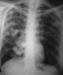
IMAGE QUIZ

Give the complete diagnosis-in this CXR-PA view
Another opportunity to win my book "Review Of Radiology".All you have to do is diagnose this case post your answers as comments here and send your full name,current occupation and postal address to sumerdoc-AT-yahoo-DOT-com.
NOTE-In case of many correct entries the prize would be given by a lucky draw.
COPYRIGHT (SUMER)
Saturday, April 9, 2005
Is infiltrate a useful term in the interpretation of chest radiographs? Physician survey results.
PURPOSE: To determine how physicians interpret the word infiltrate when it is used in a chest radiography report and if the word is helpful in the clinical management of patients.
Radiology. 2005 Apr;235(1):5-8.
Thursday, April 7, 2005
Radiation Cost of Helical High-Resolution Chest CT
OBJECTIVE. In our department, most high-resolution CT (HRCT) scans of the lungs are performed in conjunction with a standard helical examination to assess the entire chest. This requires scanning the patient twice. The goal of this study was to determine if the radiation dose could be decreased by performing a single combination helical scan of the chest from which both 5-mm standard and 1.25-mm HRCT images could be obtained.
CONCLUSION. Because the total measured radiation dose is 32% greater from a single combination helical HRCT scan of the chest versus separate standard helical plus axial HRCT scans, helical HRCT is not a clinically advisable technique.
Wednesday, April 6, 2005
Computer-aided detection (CAD) in mammography: Does it help the junior or the senior radiologist?
OBJECTIVES:: To evaluate the impact of a computer-aided detection (CAD) system on the ability of a junior and senior radiologist to detect breast cancers on mammograms, and to determine the potential of CAD as a teaching tool in mammography.
METHODS:: Hundred biopsy-proven cancers and 100 normal mammograms were randomly analyzed by a CAD system. The sensitivity (Se) and specificity (Sp) of the CAD system were calculated. In the second phase, to simulate daily practice, 110 mammograms (97 normal or with benign lesions, and 13 cancers) were examined independently by a junior and a senior radiologist, with and without CAD. Interpretations were standardized according to BI-RADS classification. Sensitivity, Specificity, positive and negative predictive values (PPV, NPV) were calculated for each session.
RESULTS:: For the senior radiologist, Se slightly improved from 76.9 to 84.6% after CAD analysis (NS) (one case of clustered microcalcifications case overlooked by the senior radiologist was detected by CAD). Sp, PPV and PNV did not change significantly. For the junior radiologist, Se improved from 61.9 to 84.6% (significant change). Three cancers overlooked by the junior radiologist were detected by CAD. Sp was unchanged.
Eur J Radiol. 2005 Apr;54(1):90-6.
Tuesday, April 5, 2005
Radiolology News-MSK ultrasound shows signs of neglect
Dr. Sandra Allison and colleagues at Thomas Jefferson University sent a questionnaire via e-mail to 300 members of the Society of Skeletal Radiology and to 105 members of the Society of Radiologists in Ultrasound. The 110 radiologists who responded (25%) had on average 13 years of experience.
More than three-quarters of respondents said that MRI should be the modality of choice to evaluate tendon, ligament, and muscle injuries. Two-thirds, however, indicated that too few musculoskeletal ultrasound examinations were ordered, and 80% of those said that reimbursement for MSK ultrasound is too low.
Almost 70% reported that their MSK ultrasound training was inadequate, while two-thirds indicated that ultrasound required too much physician time.
The survey also accounted for the specialists' respective biases. Sonologists were more likely to favor ultrasound, while musculoskeletal radiologists preferred MRI, the researchers said.