Wednesday, September 28, 2005

US in blunt abdominal trauma

Screening US and CT for blunt abdominal trauma: A retrospective study.
Marco GG, Diego S, Giulio A, Luca S
Institute of Radiology, Polytechnic University of Marche Medical School, Umberto I Hospital, Ancona, Italy.
OBJECTIVE:: To assess the accuracy of screening US and CT in patients with blunt abdominal trauma admitted to the trauma centre of our General Hospital.
RESULTS:: Screening US exhibited a satisfactory overall ability to distinguish negative from positive patients (91.5% sensitivity and 97.5% specificity in major trauma patients versus 73.3% sensitivity and 98.1% specificity in the minor trauma group) and a satisfactory specific ability to depict all injuries in major trauma patients. In minor trauma cases sensitivity was satisfactory for free fluid but unsatisfactory for organ injuries. Of the 21/864 false negative reports (5 in patients with major and 16 in cases with minor traumas), only one affected patient management, a major trauma case, by delaying an emergency laparotomy. The performance of CT in detecting each single lesion was predictably excellent in both patient groups.
CONCLUSION:: Its satisfactory accuracy for major trauma suggests that US could be employed not only to screen cases for emergency laparotomy but also as an alternative to CT. However, since major traumatic injuries generally carry an imperative indication for CT, especially as regards neurological, thoracic and skeletal evaluation, US should be employed to perform a prompt preliminary examination using a simplified technique in the emergency room simultaneously with resuscitation.
Full Article at-
Tuesday, September 27, 2005

Klatskin tumour

Radiological diagnosis of Klatskin's tumour.
Pauls S, Juchems MS, Brambs HJ
Abteilung Diagnostische Radiologie, Universitatsklinikum Ulm, .
Patients with Klatskin's tumour present clinically unspecific symptoms such as painless jaundice and cholangitic discomfort. The only curative treatment is R0 resection of the tumour. To allow stage-specific therapy, accurate tumour staging is indispensable, the first step of which is abdominal sonography. If there is a suspect finding, cross-sectional imaging techniques like MRI or MDR-CT are used to clarify or stage the lesion, respectively. To estimate resectability, MRI together with MRC and MRI angiography are superior to MDR-CT. Biopsy using ERC is required before starting any specific treatment. If therapeutically relevant peritoneal carcinosis is clinically suspected although not radiologically confirmed, PET should also be performed. The value of PET/CT has to be evaluated in further studies. For optimal treatment, close cooperation between clinicians and radiologists is necessary.
Full article at-

Saturday, September 24, 2005

Image quiz


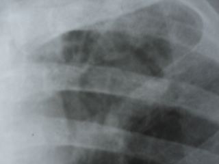
Spot the diagnosis in this CXR, leave your answers in the comments section...

Correct answers will be posted on this place after one week...

Keep coming back for more such interesting cases!!


CORRECT ANSWER-MYCETOMA (ASPERGILLOMA) IN A CAVITY (THE MENISCUS SIGN)


WINNER-Massoud Pezeshki Rad


COPYRIGHT (SUMER)

Sunday, September 18, 2005

Temporal Bone Imaging

Imaging of the temporal bone An overview.
Struffert T, Grunwald IQ, Papanagiotou P, Politi M, Roth C, Reith W
Klinik fur Diagnostische und Interventionelle Neuroradiologie, Universitatsklinikum des Saarlandes Homburg/Saar
Diseases of the temporal bone should be diagnosed by high resolution computed tomography (CT) and magnetic resonance imaging (MRI). Because of the excellent imaging of bony structures, CT is the method of choice for diagnosing pathologies of the external auditory channel, middle ear, and mastoid. Imaging of trauma is performed with CT. For examining the labyrinth system and interior auditory channel, MRI is the method of choice.
Full Article at-

Thursday, September 15, 2005

Contrast reaction-current concepts

Review article: Acute serious and fatal reactions to contrast media: our current understanding.
Morcos SK
Department of Diagnostic Imaging, Northern General Hospital, Sheffield Teaching Hospitals NHS Trust, Sheffield S5 7AU, UK.
Serious or fatal reactions to a contrast medium (CM) are unpredictable but fortunately rare. History of serious reaction to CM, bronchial asthma or multiple allergies increases the incidence of serious reactions by a factor of 5. Serious or fatal reaction to CM could be due to direct effect on basophils and mast cells or IgE mediated (type 1 hypersensitivity reaction). Activation of the kinin system leading to the formation of bradykinin could also be involved. Complement activation is unlikely to be a primary factor in initiating a serious reaction to CM. Avoiding CM administration in patients at high risk of serious reaction is advisable, but if the administration is deemed essential all precautions should be implemented and measures to treat serious reactions should be readily available. Oxygen supplementation, intravenous administration of physiological fluids and intramuscular injection of 0.5 ml adrenalin (1:1000) should be considered in the first line management of acute anaphylaxis. The ability to assess and treat serious CM reaction effectively is an essential skill that the radiologist should have and maintain.
Full article at-

Tuesday, September 13, 2005

Ultrasound in necrotizing enterocolitis

Sonographic evaluation of neonates with early-stage necrotizing enterocolitis.
Kim WY, Kim WS, Kim IO, Kwon TH, Chang W, Lee EK
Department of Radiology, CHA Hospital, Pochon CHA University College of Medicine, Seoul, South Korea.
Background: Necrotizing enterocolitis (NEC) is the most common gastrointestinal emergency in neonatal intensive care units. Ultrasonographic findings in early-stage NEC have not been described. We evaluated the echogenicity of the bowel wall, involved region, ascites, and portal venous gas at both initial and follow-up examinations.
Results: All of the neonates in the control group (n = 10) presented normal bowel wall echogenicity; the patients with NEC presented echogenic dots in 16 patients (40%) and dense granular echogenicities in 24 patients (60%). Portal venous gas was absent in all patients. On the follow-up examinations, the echogenicity of the bowel wall and ascites decreased in 37 patients (93%). The duration of NPO was 11.1 +/- 6.6 days in group I and 16.5 +/- 7.2 days in group II.
Conclusion: Echogenic dots or dense granular echogenicities in the bowel wall can be seen in patients with early-stage NEC. Bowel sonography can be helpful for the early diagnosis and monitoring of patients with NEC.
Full Article at-

Saturday, September 10, 2005

Tuberculoma versus Cysticercus granuloma-Role of Diffusion Imaging-article in Eur J Radiol

Role of diffusion weighted imaging in differentiation of intracranial tuberculoma and tuberculous abscess from cysticercus granulomas-a report of more than 100 lesions.
Gupta RK, Prakash M, Mishra AM, Husain M, Prasad KN, Husain N
Department of Radiology, Sanjay Gandhi Postgraduate Institute of Medical Sciences, MR Section, Lucknow 226014, India.
Restricted diffusion is noted in a large number of non-stroke conditions including tuberculoma. The purpose of this study was to demonstrate spectrum of diffusion weighted imaging (DWI) abnormalities in tuberculomas and tuberculous abscess and to distinguish these from degenerating neurocysticercosis. Truly T2 hyperintense lesions were divided into two subgroups, tuberculomas and tuberculous abscesses; ADC values from the core and the wall of these lesions were (0.74+/-0.13)x10(-3), (0.61+/-0.08)x10(-3) and (1.03+/-0.14)x10(-3), (1.08+/-0.14)x10(-3)mm(2)/s, respectively, and was significantly lower in core as compared to the wall. However, there was no significant difference between ADC values of the tuberculous abscess and the hyperintense tuberculomas. Vesicular and degenerating stages of cysticercus cysts from the core showed ADC values of (1.66+/-0.29)x10(-3) and (1.51+/-0.23)x10(-3)mm(2)/s, respectively, and were significantly higher than the core of all groups of tuberculomas and tuberculous abscess. We conclude that addition of DWI to routine imaging protocol may help in differentiation of tuberculous lesions from degenerating cysticercus granuloma.
Full Article at-

CT Gastrography- useful article in abdominal imaging.

CT gastrography.

Kim JH, Park SH, Hong HS, Auh YH.

The rapid dissemination of multidetector-row computed tomographic (CT) technology will make faster and more accurate gastric imagining available. Two-dimensional multiplanar reconstruction and CT gastrography including virtual gastroscopy and transparency rendering using volume rendering are types of interactive two- and three-dimensional medical imaging tools. It provides multiplanar cross-sectional imaging, gastroscopic viewing, and upper gastrointestinal series imaging in the same data acquisition. Two-dimensional multiplanar reconstruction provides accurate staging of gastric cancer and extraluminal information such as lymphadenopathy and distant metastasis. Virtual gastroscopy detects subtle mucosal changes and differentiates them from submucosal lesions in the same way as gastroscopy. Transparency rendering provides global orientation of the focal findings in the stomach in the same way as upper gastrointestinal series findings and provides useful information for preoperative mapping. Thus, CT gastrography is a promising method for evaluating gastric lesion despite its limitations.
Full Article at-

Tuesday, September 6, 2005

Sumer's Radiology Site, www.indianradiology.com celebrates successful One year

Your Favourite online Radiology Magazine celebrates one successful year..... Thanks to all visitors and Readers for their continued support.....


Image hosted by Photobucket.com