Friday, September 29, 2006

Come On Oprah! #235? You Are Soooooo Poor!

Forbes Magazine has been ranking the 400 Richest Americans since way back in 1982. In that year's issue only 13 of the mighty 400 were billionaires, the rest had merely hundreds of millions of dollars.

The 2006 issue came out last week. How many of the 400 are now worth a billion or more? The answer, after this....

Want to hear something interesting, well I think it's interesting....Flavor Flav has five children....their names are Shanique, 13; William Jr., 12; Karren, 11; Da'Zyna, 7; Quanah, 5; Kayla, 3.

Colleen, if we have a daughter....I want to name her Da'Zyna......please?

Jerry...you are wrong! Something interesting did happen in hockey this week.... The New York Islanders signed goalie Rick DiPietro to a 15-year contract where he will be making $4.75 million a year, for a total of $67.5 million! Talk about job security. His contract ends in 2021! He's going to be a 40-year old goalie...

Did you know that Rob Zombie got his start in show business as a production assistant on the Pee Wee's Playhouse?

Ok... Here's your answer....This year's Forbes 400 list of the richest Americans contains 400 billionaires for the first time! 999 million dollars doesn't even get you in the issue this year. Bill Gates is still number one with $53 billion....George Lucas was #70 with $3.6 billion, Steven Spielberg and Donald Trump were tied at #94 with $2.9 billion. Oprah is #235 with $1.4 billion....Martha Stewart dropped off the list this year. Poor poor Martha.

Thursday, September 28, 2006

Fibrous dysplasia (FD) of the skull


Findings

There is expansile lesion involving predominantly the left frontal and sphenoid bones (Figure 1 and Figure 2). The diploic space is widened with displacement of the outer table. This gives a “ground-glass” appearance with small lytic areas (Figure 1) dispersed within. This expansile, predominantly sclerotic lesion is seen to involve the sphenoid and ethmoid bones also (Figure 2 and Figure 3). The nasal cavity is distorted.
The bilateral anterior clinoid processes are involved by this expansile lesion (Figure 2), with mild narrowing of the bilateral optic canals (Figure 2).
No significant soft tissue mass is seen to be associated with the expansile bony lesion.
Figure 4: 3D rendering nicely demonstrates the abnormally deformed calvarium in this patient.


Diagnosis: Fibrous dysplasia (FD) of the skull


FD is an acquired and benign disorder in which the normal medullary space of an affected bone is replaced by fibrous tissue. The disorder usually manifests in childhood and adolescent years, and presents as either an incidental finding in asymptomatic patients or with nonspecific symptoms, such as pain, swelling, or tenderness. Skull involvement can lead to hearing impairment. There are monostotic (involving a single bone) and polyostotic (involving multiple bones) forms. The monostotic form is much more prevalent than the polyostotic form. FD can affect any bone; however. when solitary, the most common bone to be affected is the proximal femur, followed by the ribs and craniofacial bones. The polyostotic form can vary, and usually affects the femur, tibia, pelvis, and bones of the feet. The association of polyostotic fibrous dysplasia with precocious puberty in girls, and cafe-au-lait spots is termed the McCune-Albright syndrome. The combination of intramuscular myxomas and polyostotic fibrous dysplasia is called Mazabraud syndrome.

Because FD is a medullary process, the radiographic appearance consists of a lucency with an epicenter that is intramedullary or eccentric, but not cortical. The lesion can appear cystic and expansile with endosteal scalloping, but is always well defined without breaking through the bony cortex. The lucent lesion is usually described as having a ground-glass quality. Another classic appearance includes the shepherd’s crook deformity, which describes the appearance of the femoral neck that results when the disorder causes varus angulation. FD of the calvarium appears as increased density, usually at the skull base. Paget’s disease can give this appearance as well; Paget’s is generally bilateral, whereas FD is unilateral.

There is rare potential for malignant transformation, especially in the polyostotic form. When radiologic features such as cortical destruction, periosteal reaction, or a soft tissue mass are present at the site of prior FD, malignant degeneration should be suspected. Progression to osteosarcomas, fibrosarcomas, and malignant fibrous histiocytomas has been reported.

Most lesions appear low to intermediate signal on T1-weighted MR images. The signal varies on T2-weighted images, but is generally high in signal, especially with the presence of nonmineralized and cystic components. Enhancement is present, and is usually heterogeneous. Because the MR imaging appearance is variable and can imitate neoplasm, correlation with plain films or CT is helpful when fibrous dysplasia is suspected. MRI is useful for evaluating cranial neuropathy and suspected malignant transformation.

CT shows three overlapping patterns - pagetoid (mixed) pattern in 50%, sclerotic pattern in 25%, and cystic pattern in another 25% of cases. The lesions usually show nonspecific increased uptake of radiotracer on bone scans.

Involvement of the skull may cause cranial nerve dysfunction with visual and hearing impairment. Mucoceles may complicate paranasal sinus ostial bone involvement. The term "leontiasis ossea" describes a rare form of polyostotic disease that involves the frontal and facial bones. Another craniofacial entity, “cherubism,” is a hereditary fibrous lesion of bone, symmetrically involving the mandible and often the maxilla.

I'm Going To Write About Sports - Why? - Because I Have A Penis - That's Why!


Do you believe in the Madden Curse? Electronic Arts executives announced Seattle Seahawks Running Back Shaun Alexander would be on the cover of the 2007 version of the Madden Franchise. It was recently announced that Alexander would be out indefinitely with a broken foot. Now it turns out that it will be approximately two to three weeks before Alexander can return to the team.

Detroit Lions Running Back Barry Sanders - Sanders appears on the 2000 edition of the Madden Franchise. Known as one of the best running backs in NFL history, Barry Sanders abruptly retired one week before training camp started for the season. At the height of his career, Sanders retired just 1,500 yards short of becoming the NFL's All-Time Leader in Rushing Yards.

Tennessee Titans Running Back Eddie George - George appears on the 2001 edition of the Madden Franchise. The previous year, George led his team to the Super Bowl. George had a career season that year, however, he bobbled a pass in the playoffs that year, which led to a turnover, which was returned for a touchdown, which cost the Titans the game. The following season, his rushing average sank to an all-time low.

Minnesota Vikings Quarterback Daunte Culpepper - Culpepper appears on the 2002 edition of the Madden Franchise. The previous year he had led his team to the NFC championship game. The year he was on the cover, the Vikings struggled to a 4-7 record, until Culpepper was shelved with a season-ending knee injury.

St. Louis Rams Running Back Marshall Faulk - Faulk appears on the 2003 edition of the Madden Franchise. Faulk led the St. Louis Rams to two Super Bowls in the three previous years, and had more than 1,300 yards rushing in each of those seasons, before appearing on the cover of the Madden football game. The year Faulk was on the cover, he suffered an ankle injury, dropped to under 1,000 yards rushing, and had a bad season overall. He never broke through the 1,000 yards rushing mark for the rest of his career.

Atlanta Falcons Quarterback Michael Vick - Vick appears on the 2004 edition of the Madden Franchise. Vick fractured his right fibula in a preseason game ONE DAY after the game hit store shelves. Vick played in only 5 regular season games, finishing with only 585 yards passing and 4 touchdowns. The Atlanta Falcons missed the playoffs, finishing the season with 5 wins and 11 losses, a stark contrast on the team's 9-6-1 record the season prior.

Baltimore Ravens Linebacker Ray Lewis - Lewis appears on the 2005 edition of the Madden Franchise. Lewis suffered an injury that kept him out of the last game of the regular season. It was also Lewis' first season without a single interception, after posting a career-high 6 the previous year. The Baltimore Ravens also failed to make the playoffs that season, after winning their division the year before. Furthermore, in Week 6 of the following year, Ray Lewis' season was ended by a torn hamstring.

Philadelphia Eagles Quarterback Donovan McNabb - McNabb appears on the 2006 edition of the Madden Franchise. In the first game of the regular season, McNabb took a hit to the chest from the helmet of Atlanta Falcons defensive tackle Chad Lavalais. After the game, it was revealed that McNabb had a 'sports hernia' that needed surgery. Refusing to have the required season-ending surgery while the Eagles (who had made the Super Bowl the previous season) still had a chance to make the playoffs, McNabb played in discomfort and pain throughout the season. On November 21, 2005, McNabb, on the advice of his doctors, opted to have season-ending surgery for his sports hernia; the Eagles had been eliminated from playoff contention and finished the year with a 6-10 record.

Madden is on the cover of the Hall of Fame edition of the game this year. Will he make it through the 2006-2007 season? We're going to have to wait and see.

Tuesday, September 26, 2006

How To Interpret a CXR-Good Article

Very Interesting Full article here- Looking at the chest radiograph, From London South Bank University

It goes on like-

"The good radiologist has been trained to seek those features that normally lie outside of the awareness of regular folk in the real world. Originally the human visual system was evolved to enable us to make very fast assessments and then to get out of the way of danger that was seeking us. The processing involves compromises that are not always appropriate to Radiological assessment. Most approaches to the teaching of radiographic interpretation involve a combination of systematic analysis and looking for remembered patterns. The good Radiologist has to be consistent and training involves at least one imposed scheme. It may be boring to go through a list each time but initial ennui saves money in later indemnity charges.

For each radiographic projection, the beginner is advised to have a list of features that they will deliberately observe. Repetition will convert this to an automatic response, rather similar to the concert pianist who does not have to be deliberately aware of fingering, but can concentrate on musical interpretation. " Full Article here

Monday, September 25, 2006

Radiology Grand Rounds-IV

Welcome to the Fourth Edition of Radiology Grand Rounds, Posted late because of some problems with the Scan Man's Blog, the original host for this edition of Radiology Grand Rounds.
This edition will be divided into sections in same way as is done in Radiology Journals.
Sections
  • Pediatric Radiology
  • Chest Radiology
  • Interventional Radiology
  • Radiation Physics
  • Computers in Radiology
  • Editorial-Radiology Malpractice

PEDIATRIC RADIOLOGY
Clark Bartram of Unintelligent Design shares with us a case of Transient Tachypnea of the Newborn with a good image & good explanation of TTN. It also has story that goes with this account of TTN and the discussion as to why this is more common in babies who have been born through a C-section.

Dr Sethi points towards Pediatric aspects of radiation exposure A briefing on CT radiation exposure and some radiation hazards what all pediatricians should know.

CHEST RADIOLOGY
Signs in Chest Radiology
"The classic radiographic finding of pulmonary infarction is a wedge-shaped, pleural based triangular opacity with an apex pointing toward the hilus (Hampton hump)."

Another Interesting Chest X-ray by FilmJacket.com, try and make the diagnosis in the radiograph before you read the entire case.

INTERVENTIONAL RADIOLOGY
Dr. Bhavin's blog features a case of Radiofrequency ablation of a peripheral lung tumor in a 78-year-old man with emphysematous lungs.

RADIATION PHYSICS
Sumer's Radiology Site talks about a basic radiation physics concept for medical students with a funny illustration.

COMPUTER IN RADIOLOGY
So you bought a new computer, took it out of the box and positioned it on your desk. A short while later, you realized that without the right software, your computer is nothing but an expensive brick. Then the reality set in - you have to pay several hundred dollars for an office suite such as MS Office, another few hundred bucks for a good image-editing package such as Adobe Photoshop, and that's just the beginning. You may need good encryption software, an antivirus software, a firewall, PDF creation software and, if you are like me, software to help you establish web presence. Before you know it, you could be out more than a thousand dollars.
Or not! There is a way to equip your computer with all the tools you need, using high quality, full-featured and stable software that won't cost you a penny. I have been using the following open-source software for a long time and have been extremely pleased not only with software performance, but with all the extra money in my pocket. READ MORE

EDITORIAL

How to avoid medical malpractice litigation

If, as a radiologist, you are concerned about the possibility of medical malpractice litigation, you are not alone. Almost everyone has heard of at least one colleague getting sued. Malpractice insurance premiums are sky-high. Malpractice lawyers are waiting to pounce on any case that looks promising. The consequences of successful litigation can be very profound. However, there are certain simple things that may help avoid successful malpractice litigation against you. READ THE FULL ARTICLE

That wraps up this month's highlights of the Radiology blogosphere. Hope the readers enjoyed the fourth edition of the Radiology Grand Rounds. If you liked any of these blogs, keep visiting them. Please email me at sumerdoc@yahoo.com if you are interested in hosting future Radiology Grand Rounds. Archive for the Radiology Grand Rounds here-Radiology Grand Rounds.For More updates on Radiology Grand Rounds A new discussion Grouphas been created here, send me a mail to be invited to the group.Group name: Radiology Grand RoundsGroup home page: http://groups.google.co.in/group/radgrandroundsGroup email address radgrandrounds@googlegroups.comBe sure to tune in Next Month Last Sunday 29 th October when the Radiology Grand Rounds will be hosted by me again at Sumer's Radiology Site, email your submissions here- sumerdoc@yahoo.com

The Cathedral of Sts. Peter and Paul, Calbayog City, Western Samar

Click on image to enlarge.

Calbayog City at Dusk

Shot near the Calbayog Market on Navarro street on the eve of the town fiesta. Sept. 7, 2006. Click on picture to enlarge the image.

Sunday, September 24, 2006

Bizzare Radiograph

Amazing what all you can discover on an abdominal Radiograph these days
Read this-
Delicate Situation

( Hat Tip-Grunt Doc-Must-see X-ray; also, a foreign-body tale )

Radiology Residency Interview-Do & More importantly Donts'

Found this on FilmJacket.com, very interesting..
"There is plenty of information out there on how to excel during a residency interview. Or is there? More specifically, is there any information online or in the books on how to ace the radiology residency interview? Instead, we are going to tell you how to screw up with flare and style. If you are really astute, you may figure out what not to do!"
Read the full article here-

Pediatric Grand Rounds Are Up


Paediatrics Grand Rounds Are up on www.pediatricsinfo.com

Here-Pediatrics GR

:) Yours Truly Features in it!!

Saturday, September 23, 2006

Calbayog Sights

Rebelito's. This is one of the oldest refreshment stops in Calbayog City. Founded in the 1950's, Rebelito's was originally built as a simple "Halo-halo" store built like a "Bahay-Kubo". The specialty is still the Halo-halo(which is a concoction of tropical fuits and deserts mixed in crushed ice and milk.
Calbayog Bakery along Magsaysay Boulevard


Entrance to the Fish Port. It's always good to shop for seafoods in this place, the prices are cheap and the seafoods fresh. I always look around every morning to look for tuna and squids.


A village in the Fish Port


San Joaquin Inn and Restaurant in front of the town market, along Nijaga Street. 600 pesos for 24hours' stay at one of their airconditioned rooms, with private toilet and cold and hot shower(no cable Tv though). I stayed here for three days.

For those on tight budget, they offer rooms for only 350pesos but no aircondition or TV set. There is no room toilet so you share a common toilet with other tenants. Open 24 hours.

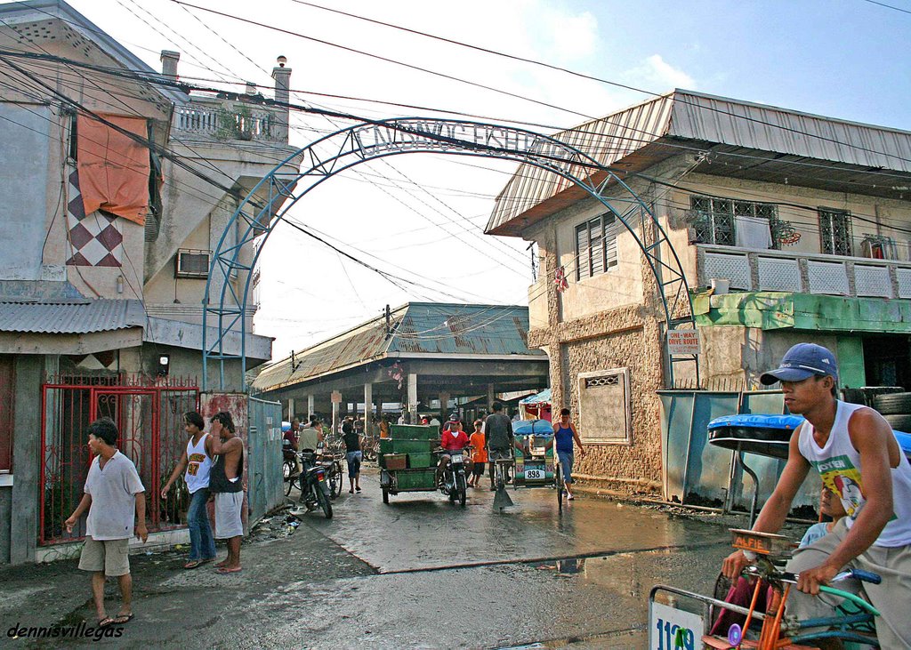
Eduardo's Hotel, along Pajarito street. I stayed here during my first trip to Calbayog way back in 2004. This hotel is the best there is in Calbayog. They charge 700 pesos for a double-bed with aircondition, Cable TV, and hot and cold shower, with the benefit of ordering your food anytime day or night.

MORE PHOTOS SOON...

Friday, September 22, 2006

"Writely" - An Online Word Processor

All gmail users can now use writely. This means that even if you have never signed up for it, if you have a google account you now can.
http://www.writely.com

With Writely, you can:
Use our online editor to format documents, spell-check and more.
Upload Word documents, OpenOffice, RTF, HTML or text.
Download documents to your desktop as Word, PDF and more.
View your documents' revision history and roll back to any version.


Plus, since its online, you can:
Invite others to share your documents by e-mail address.
Edit documents online with whomever you choose.
Publish documents online to the world, or to just who you choose.
Post your documents to your blog.

Grilled

I watched a great movie last night! If you haven't seen the movie "Grilled", go rent it! I don't know how this movie slipped through the cracks, but I did not hear and advertising for the movie. The movie stars Ray Romano, Kevin James, Michael Rappaport, Burt Reynolds (who now officially looks like a piece of beef jerky), Juliette Lewis, and Sophia Vergarra (ding ding ding!) Kevin James and Ray Romano play struggling meat salesman who get caught up in organized crime. I know it sounds horrible, but it was really good. If you are a fan of the dark comedy, go rent this movie!

Madonna has set the record for highest grossing tour for a female artist. Tour producer Arthur Fogel puts the tally at $193.7 million from 60 shows that drew nearly 1.2 million in paid attendance.

That gross narrowly puts Madonna over Cher's benchmark of $192.5 million. However, it took Cher an endurance-testing 273 shows between June 2002 and April 2005 to reach that mark on her marathon Farewell tour.

Oh look! Madonna on a cross! My blog is soooo controversial.
Can't you just go on a Tuesday morning? Six Flags Great America is using new tactics, other than the creepy dancing old guy, to try to entice people to visit their theme parks. Cockroaches. Starting October 7, 2006, the Six Flags theme park in Illinois will offer park visitors a pass for four people to cut to the front of ride lines. The stipulation? They will have to eat a live 2- to 3-inch horned Madagascar hissing cockroach.

The park will also offer a cooked roach eating contest to park visitors on Friday the Thirteenth of October. And in case you wanted to know....The world record for eating cockroaches was set by a British man who ate 36 cockroaches!

Thursday, September 21, 2006

Pyogenic cerebral abscess


Findings

There is a well-defined, smoothly marginated, thinly rim-enhancing intraaxial mass (Figure 4) with surrounding edema (Figure 3) and mass effect centered in the right deep gray matter. The rim is essentially isointense to white matter on the T1-weighted image (Figure 1), hypointense on the T2-weighted image (Figure 2), and there is homogeneously increased signal intensity of the mass on the diffusion-weighted image (Figure 5).


Diagnosis: Pyogenic cerebral abscess


Most pyogenic cerebral abscesses are located supratentorially at the gray - white junction, secondary to hematogenous spread, and within the frontal and parietal lobes (middle cerebral artery distribution). Size is variable and may range from a few millimeters to many centimeters.

The characteristic imaging findings are illustrated in this case, but the pathogenesis and assoiciated imaging findings have been divided into four stages, which have been extensively published and are as follows:
- early cerebritis
- late cerebritis
- early capsule formation
- late capsule formation

Although hematogenous dissemination from an extracranial infection is the most common source, other potential etiologies to consider include: hematogenous spread from any right-to-left shunt, penetrating trauma, postoperative, and direct extension from a paranasal sinus, meningeal, calvarial, or otic infection.

These relatively uncommon lesions are potentially fatal but treatable, with treatment (often depending of the size of the lesion) typically ranging between systemic antibiotics alone and stereotactic aspiration or drainage. Reports of mortality vary, with published numbers between 0% and 30%. It is important to be aware of the complications of incompletely or inadequately treated abscesses and to look for them on MR.

These include:
- Intraventricular rupture with ventriculitis
- Meningitis with satellite abscess development
- Hydrocephalus
- Local mass effect or herniation.

Wednesday, September 20, 2006

Link to Vascular Atlas


3D Vascular Atlas with Volume Rendering is available at-3D Vascular Atlas
Authored by Frank Corl, Melissa Garland, Brian Kuszyk, and Elliot Fishman.
The educative material has separate sections on Syllabus, Lectures, Related Protocols, Selected Readings and Case Studies of Head and Neck, Thoracic and Abdominal Syllabus, Lectures, Related Protocols, Selected Readings and Case Studies of Head and Neck, Thoracic and Abdominal regions.

Tuesday, September 19, 2006

Having Fun with Radiology

Found this interesting link while surfing the net, Interesting Radiographs making Radiology Look interesting-

FUN WITH RADIOLOGY

Kickball

Does anyone want to join a kickball team with me? Chelsea? Jerry? Any of you that read my blog? They play in Irvine..... I don't want to join by myself.... Anybody?

Come on....You owe me.... Why, you ask? Nobody told me that the United States now has an elephant polo team! There was a posting on CraigsList that included:

Join the DC Elephant Polo Team as we go to Thailand for the 2006 Kings Cup Elephant Polo Championships! . . . No previous polo experience is necessary.

They just went to Hua Hin, Thailand, where they played polo on elephants for the first time! I am so jealous!

The practices consisted of the teammates batting a ball around and waving homemade eight-foot-long bamboo mallets - while standing on part of a swing set and an electrical utility box to simulate an elephant's height.

So, how about it? Kickball anyone?

ARRRRRRRRRRRRRR!!!!!


Monday, September 18, 2006

TRIP database is now free access

TRIP Free Access Evidence-Based Medicine Database - Turning Research Into Practice
http://www.tripdatabase.com/index.html

TRIP Free Access Database is one of the Internet's leading resources for Evidence-Based Medicine. Allowing users to easily and rapidly identify the highest quality evidence from a wide range of sources. The TRIP Database began in 1997 as a result of the work of the founders (Jon Brassey and Dr Chris Price) in answering clinical questions. The TRIP Database is produced by TRIP Database Ltd. For approximately 4 years the TRIP Database became a subscription service which ended in September 2006. This allowed for a dramatic improvements in the site. However, it also significantly restricted use. The TRIP Database is now free access and truly a great resource to the Internet clinical research community.

Neurology Image Library

Just discovered this excellent collection of Radiological Images of Neurological cases. This collection is maintained and copyrighted by-

Internet Stroke Center at Washington University

(The Internet Stroke center is an independent resource about stroke care and research.)

Hypoxic ischemic encephalopathy


Findings

Figure 1 and Figure 2: Axial CT images show linear high attenuation subdural collections consistent with hematomas. No fractures were seen on bone windows.
Figure 3: Axial CT image shows an extracalvarial hematoma.
Figure 4, Figure 5, Figure 6, Figure 7, and Figure 8: DWI shows high signal demonstrating restricted diffusion in the thalami bilaterally (Figure 4). High signal is seen in the lateral temporal parietal regions (Figure 5), and the splenium of the corpos callosum (Figure 6), as well as the body of the corpus callosum (Figure 7), and the high parietal regions (Figure 8).


Diagnosis: Hypoxic ischemic encephalopathy


Hypoxic ischemic injury can present in a variety of patterns, including:
- Periventricular leukomalacia (PVL)
- Cortical watershed ischemic changes
- Deep gray matter ischemia
- Mixed pattern

Hypoxic ischemic injury results from a global hypoxic or ischemic insult to the brain in utero or during birth. Factors that can contribute to neonatal brain injury include placental abruption, meconium aspiration, cord prolapse, dystocia, eclampsia, maternal hypotension, and severe maternal bleeding.

Various patterns of brain injury have been described based upon pathological specimens.

They include:
(1) PVL that does not involve the cortex,
(2) injury to the parasagittal cerebral cortex
(3) involvement of the deep gray matter
(4) mixed injury pattern, which may or may not include hemorrhage.

Because the brain injury is global, the patterns of brain injury are, in general, bilaterally symmetric.

The imaging evaluation of neonates in whom a hypoxic injury is suspected, can include a variety of modalities including ultrasound, CT, and MRI. Ultrasound is a useful modality to detect PVL, which usually affects premature infants. PVL results in response to ischemia in the watershed areas of the brain, and presents as areas of increased white matter echogenicity, predominantly in a posterior distribution. The affected white matter can eventually develop small cystic cavities and, in severe cases, result in atrophy and ventriculomegaly. These findings may be evident on CT and MRI as well.

Whether there is involvement of the cortex or the deep gray matter, usually depends on the degree of the hypoxic insult. Severe injury tends to affect the deep gray structures, including the basal ganglia and thalami, which can show bright T1 weighted signal. Less severe injury will lead to ischemic changes in the cortical watershed areas. These affected areas will also show restricted diffusion. The corresponding areas are hypodense on CT, secondary to the presence of edema.