Friday, November 30, 2007

Funny Religion Joke

Three men arrive at the gates of heaven. St. Peter asks the first man,

"Religion?"

The first man replies, "Episcopalian."

St. Peter looks down his list and says, "go to room 24. But be very quiet as you pass room 8."

To the second man St. Peter asks, "Religion."

The second man replies "Methodist."

St. Peter looks down his list and says, "Go to room 14. But be very quiet as you pass room 8."

To the third man St. Peter asks. "Religion."

The third man replies, "Baptist."

St. Peter looks down his list and says. "Go to room 21. But be very quiet as you pass room 8."

The third man then says to St. Peter, "I can understand there being different rooms for different religions, but why must we be quiet when we pass room 8?"

St. Peter tells him, "Well, the Catholics are in room 8, and they think they're the only ones here."

Penis Cutting

Ok....so....hmmm..... Do you know about Kenyan Tribe circumcision rituals?

They usually have a ceremony when the boy turns 14 or 15. The boy is circumcised in front of the tribe, and if the boy flinches he is apparently considered a disgrace to his family. Sweet.....

Thursday, November 29, 2007

Strolling in Cubao Before Curfew Time

Before Curfew hour (12-5a.m), I decided to take a stroll in Cubao to take pictures of the recently unveiled giant Christmas tree and the spectacular lighted trees in Araneta Center.

It was already 10:30 in the evening, and I have to take pictures fast, or I will end up being arrested and spending the night in jail (although it would have been quite an experience to spend a night in jail).

And so here they are, a few portraits of my Cubao stroll--prior to the arrival of the police to enforce Curfew.

The behemoth Christmas Tree of Cubao


The butterfly Christmas decors that lined the streets of Araneta Center


The Gateway Mall


Ali Mall, blanketed by Christmas lights


The beautiful lighted trees of Cubao


The giant Christmas Tree at close range

Dacryocystitis


Findings

Ring enhancing lesion in the region of the right lacrimal sac, contiguous with the nasolacrimal duct. Surrounding preseptal edema. Scattered relatively mild ethmoid air cell mucosal thickening and bilateral maxillary sinus mucosal thickening. Bones are unremarkable. A mucous retention cyst is seen in the nasopharynx.

Clinical differential diagnosis:
- Cellulitis
- Abscess
- Dacryocystitis


Diagnosis: Dacryocystitis


Stagnation of tears in a pathologically closed lacrimal drainage system can result in dacryocystitis.
Acquired dacryocystitis can be acute or chronic.
There is also a form of congenital dacryocystitis, thought to be related to lacrimal excretory system embryogenesis, specifically incomplete canalization of the nasolacrimal duct.
Acute dacryocystitis is manifested by the sudden onset of pain, erythema, and edema overlying the lacrimal sac region.
Complications include abscess formation and spread of infection into the adjacent orbit.

Wednesday, November 28, 2007

Medulloblastoma


Findings

Large solid and cystic midline heterogeneously enhancing cerebellar mass effacing the fourth ventricle and causing lateral and third marked ventriculomegaly with transependymal edema. Two foci of leptomeningeal contrast enhancement in the posterior fossa, possibly metastases.


Differential Diagnosis:
- Pilocytic astrocytoma
- Medulloblastoma
- Ependymoma
- Brainstem glioma


Diagnosis: Medulloblastoma, undifferentiated type (WHO Grade IV)


Discussion

Medulloblastoma is somewhat more common in males than females with a ratio of approximately 1.5 to 1. This tumor accounts for approximately 7-8% of all intracranial tumors but over 30% of pediatric brain tumors. 75% of the cases occur in children, but the tumor can be seen at any age. The median age of presentation is 9 years. Presentation usually is secondary to hydrocephalus which develops because of mass effect from the tumor on the normal cerebrospinal fluid pathway. Truncal ataxia and trouble walking can be seen secondary to tumor invasion in to the cerebellum. Medulloblastomas can be seen in syndromes such as Gorlin's and Turcot's syndromes.

Medulloblastoma is a primitive neuroectodermal tumor which means it is a small round blue cell tumor. These types of tumors typically are tightly packed which results in a higher density than surrounding soft tissues. This fact helps with the diagnosis as these tumors will appear dense on noncontrasted head CT. This tumor will typically intensely and homogenously enhance. Calcification can be seen but it is uncommon.

Medulloblastoma can become metastatic via CSF spread. This a poor prognostic factor. Preoperative MRI can help identify metastatic lesion so they can be removed at initial surgery if possible. Surgery is the first line therapy for medulloblastoma. Postoperative chemotherapy and radiation are employed as well. The radiation therapy is extensive with the entire spine and brain being irradiated. With the combination of these three therapies the patients can reach an 80% 5-year survival rate excluding the metastatic at time of diagnosis patients.

Wednesday, November 21, 2007

I Am Not a Bibliophile

I was browsing through a pile of bargain books in Booksale last night when I came across the book The Little Prince by the French author Antoine de St. Exupery. It was tagged 70 pesos, and I thought that it was really a steal. My first impulse was to buy it right away--despite the fact that I already read it a hundred times and own at least five copies of it stuck away somewhere in my library.

But I didn't. Instead, I placed it on a prominent display for other people to easily spot it. The book is such a gem that I thought it should be required reading in all schools--elementary or post-graduate. If you haven't read it yet, go and buy a copy. And let me know your thoughts as well.

Anyway, the topic of books gives me a lot of things to talk about--because I'm a rabid reader. I read everything, from classics to trashics--well, I even read the telephone directory once in a while. Reading numbers will give you a sort of relaxation after hours of reading all words.

But then, there exist books in my library that made me feel like I'm a literary genius.

Like James Joyce's Finnegan's Wake.

Well, I survived reading James Joyce's Finnegan's Wake. Only to be hospitalized a week later for total exhaustion. My doctor told me not to read the book again, or it would be fatal next time. Now don't force me to give the summary of Finnegan's Wake--or I'll be force to give it. Okay, you really want the plot, eh?

Here it is:

Beddings makeith jared Finnegan higwiga frilles-in-pleurs two-gamers havest likened thou meest senet gorgios mumper of a once walstrait oldparr offwall webbingstone upturnpikepointandplace is at the knock out likely are we are we. Quick lunch by our left, whee. But fahr, be fear! And enthewsyass cuckling a hoyden to go the New Livius Lane where we whiled while we whithered. Conjugarly Finnegan woke up enthesyway whee.

You didn't understand the plot? Well then, go ahead and read the book yourself and see if you can even understand the first two pages. No wonder Mr. Joyce spent a lot of times in the hospital trying to finish the book. It could have most likely killed him, since it was his last novel. But don't be misled by me. The book has been highly-acclaimed as one of the world's literary masterpieces. Ho-hum...

Also one of my favorite books is the Da Vinci Code--although I haven't read it yet. In fact, it's still sealed in the original plastic wrap the way I bought it brand new from National Bookstore. I've just watched the movie version and I think I liked it. So I didn't bother reading the book. Anyway, I plan to read it someday, when I get tired of watching the movie.

I also had the courage to read Stephen Hawking's excellent A Brief History of Time, a book I couldn't read past page five. It was excellently written, of course. It's me who is at fault for not being able to understand beyond page five. And hats off to Mr. Hawking for including only one mathematical equation in his book, Einstein's famous E=mc2 . Until now, I have yet to know the meaning of that equation. All I know is that it invented the atomic bomb.

Around my house from kitchen to living room, and well into the bathroom and portions of the roof, you will find stacks of books on virtually every topic: History, Literature, Biography, Religion, Sex, Politics, etc. I read each one of them....if I could find the time to read them. Some books take just a few hours to read, and some a few years.

Some several years.

For example, I am now in my tenth year trying to read Arnold Toynbee's A Study of History, and I think it will take me ten more years to complete reading it. Its a monumental work, indeed, and must take monumental time to read. I plan to auction the book in Ebay after reading it. Or else it would just gather dust in my bookshelf.

Others only take one sitting to read: like Hartzell Spence' Marcos of the Philippines. You can read a few chapters and then throw it away, or better yet, make origami from it. The pages can also be useful as pambalot ng tinapa.

Or perhaps Adolf Hitler's Mein Kampf. Yes, I'm maybe one of the few Filipinos who dared read the book. Actually, I bought it from a friend, who later had to be treated for schizophrenia days after reading the book. I confess, reading the book is like putting a live socket in your nose and getting an electric jolt in your brain. It will make you lethargic for days.

Many years ago while a student in Manila, I bought what was probably one of the greatest books ever written: Albert Einstein's Special Theory of Relativity. Well, my reason for buying the book is not really to read it.

I just thought it was cool to carry the book around and let people be amazed that I was reading Einstein. I even bought fake eyeglasses to complement my act of playing genius. Inside the jeepney, I would feign reading it, and people would look at me reading all those formulas and equations, thinking I'm a genius. Truth is, I didn't understand the book.

Not a word of it.

Actually, according to surveys, only three people actually understood the book. One of them was Einstein.

I have read more books than I can afford to share here. But then, I'm afraid you might think I'm one of those guys wearing buttoned-up long-sleeves with suspenders and ultra-polished hair and shoes. I may look nerd but I'm not a nerd.

I also accept book donations, if you want me to review your favorite books. Right now, I'm still busy reviewing Plato's Republic, Immanuel Kant's Metaphysics, and Boswell's Life of Johnson. I hope to survive reading these books in order to continue with Arthur Hailey's Airport, Sun Tzu's Art of War, Solzhenitsyn's Gulag Archipelago, and Erica Jong's Fear of Flying.

And I also plan to read the Qu'ran someday--in original Arabic....

Meantime, I'm off to read The Little Prince for the 101st time.

Radiology Grand Rounds XVIII


Here is a case of Acetabular fracture for the Radiology Grand Rounds submitted by Dr MGK Murthy, Dr Sumer Sethi of Teleradiology Providers. Concept and Archive of the Radiology Grand Rounds is available at- Radiology Grand Rounds.


This patient had posterior dislocation of hip reduced about 6 months back now he presents with complaint of pain in the left hip.

FAQ (questions to be answered)
(a)Is it unreduced?
NO

(b) Has it developed AVN?
NO

(c) Is there any associated injury which Xray did not pickup?
MRI is silent on it


(d) Can anything else help?
YES CT would help

(e) What does it show?
It shows post wall fracture comminuted with loose fragment in the joint cavity

(f)What are the complications of posterior dislocation hip?
Complications-- include avascular necrosis, osteoarthosis, sciatic nerve injury and heterotrophic ossification.

(g) What are the types of acetabular fractures?
Acetabular fractures are classified according to Judet classification usually.
Walls, Columns and Transverse varieties


Wall fractures
Anterior wall
Posterior wall
Posterior column with posterior wall (also a column fracture)
Transverse with posterior wall (also a transverse fracture)


Column fractures
Anterior column
Posterior column
Both-column
Posterior column with posterior wall (also a wall fracture)
Anterior column with posterior hemitransverse (also a transverse fracture)


Transverse fractures
Transverse
T-shaped
Transverse with posterior wall (also a wall fracture)
Anterior column with posterior hemitransverse (also a column fracture)


Common types (90%)
Both-column
Transverse with posterior wall
Posterior wall
T-shaped
Transverse


Hope you enjoyed this edition of Radiology Grand Rounds submissions are requested for the next Radiology Grand Rounds posted every month last sunday. If you interested in hosting any of the future issues contact me at sumerdoc-AT-yahoo-DOT-com.

Dural sinus thrombosis, with involvement of the superior sagittal sinus, straight sinus, and vein of galen


Findings

CT: There is marked dilation of the superior sagittal sinus with low attenuating density, containing high density septations. The expansion and density extends into the straight sinus. There is a large peripherally calcified lesion at the right aspect of the dilated superior sagittal sinus near the torcular Herophili. There is also severe hydrocephalus, with involvement of the third and lateral ventricles.
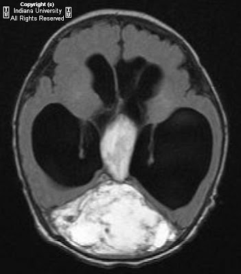
MRI: There is a heterogeneous lesion expanding the superior sagittal sinus and torcula, demonstrate mostly hyperintense signal on T1 and T2, containing multiple septations, with suggestion of fluid/fluid level, without definite contrast enhancement. There is extension into an enlarged vein of Galen. There is a area of low signal on the right side corresponding to calcification seen on CT.
No central flow is demonstrated in the posterior portion of the superior sagittal sinus, corresponding to the lesion.
Angiography (images not shown): Showed mass-effect in the posterior parietal-occipital regions without filling by contrast. No evidence of tumoral blush, AV shunting, or aneurysm.
Follow-up CT: Showed significant decrease in mixed density material expending the superior sagittal sinus from the vertex to the torcula, consistent with interval resorption of clot.

Differential diagnosis for the neonatal studies:
- Dermoid
- Vascular tumor
- Congenital variant
- Giant dural sinus thrombosis


Diagnosis: Dural sinus thrombosis, with involvement of the superior sagittal sinus, straight sinus, and vein of galen
Diagnosis confirmed on the subsequent CT, which demonstrated partial resorption of thrombus.


Discussion

Dural sinus thrombosis represents formation of clot in the dural sinus
Clot spreads from dural sinus to cortical veins, resulting in hemorrhage and infarct
Will progress to infarction in approximately 50%

Clinical presentation:
- Presenting symptoms include headache, focal neurologic deficits, papilledema
- Numerous etiologies, such as hypercoagulable state, infection, inflammation
- Dehydration and infection is a common cause in children
- Idiopathic in 10 to 30% of cases
- No particular age or sex predilection
- Morbidity and mortality from progression to venous infarction
- Treatment include thrombolysis

Imaging:
- Early findings are subtle
- Classic sign is the "empty delta" seen on contrasted imaging
- Thrombus seen in dural sinus and cortical veins
- Sinus is occluded and expands
- Appearance depends on age of clot
- Vessels may recanalize

CT:
- "Empty delta" sign represents non enhancing thrombus in the dural sinus with surrounding enhancing dura in contrast enhanced CT
- "Cord sign" is hyperdense dural sinus clot seen in non contrasted CT
- May see associated hemorrhage or edema

MRI:
- Signal on T1 and T2 depends on age of thrombus
- On GRE, the thrombus may "bloom," depending on stage of hemorrhage
- On MRV, there is absence of flow

Angiography:
- Thrombosed sinus does not fill

Hydranencephaly


Findings

Figure 1: Noncontrast CT of the head reveals complete absence of supratentorial brain tissue which is replaced by cerebrospinal fluid. The falx is present.
Figure 2: Noncontrast CT of the head reveals complete absence of supratentorial brain tissue with residual pons and cerebellum.
Figure 3: Axial T2 reveals complete absence of supratentorial brain tissue which is replaced by cerebrospinal fluid. The falx is present.
Figure 4: Axial T2 reveals complete absence of supratentorial brain tissue with residual pons and cerebellum.
Figure 5: Sagittal T1 reveals the presence of the tentorium cerebelli and structures of the midbrain along with previous findings.


Diagnosis: Hydranencephaly


Hydranencphaly is a congenital malformation of the brain in which there is near total to complete absence of the cerebral cortex and basal ganglia. There is usually preservation of the thalami, cerebral peduncles, pons, midbrain and cerebellum. Although the main etiology is unknown, it is thought to be secondary to an in-utero event. Possibilities include infection (CMV, HSV, Toxoplasmosis), vascular accident, specifically occlusion of the anterior circulation, or diffuse hypoxic-ischemic necrosis due to maternal exposure to carbon monoxide or butane gas.

Hydranencephaly can be difficult to distinguish from extreme hydrocephalus, alobar holoprosencephaly and porencephaly. The presence of residual cerebral cortex helps to distinguish hydranencephaly from the other diagnoses. The absence of the third ventricle and presence of the falx also aids in differentiation since the third ventricle is present in the other diagnoses and the falx is absent in holoprosencephaly.

In utero ultrasound is helpful in diagnosing hydranencephaly. This usually presents as a large cystic mass filling the entire cranial cavity with absence or discontinuity of the cerebral cortex. The appearance of the thalami and brainstem protruding inside a cystic cavity is also characteristic. CT and MRI findings are similar with the absence of most supratentorial structures and preservation of the brainstem and cerebellum. MRI is superior for the detection of cortical remnants.

Prognosis in these children is very poor with most deaths occurring within the first year. Although children with hydranencephaly may appear healthy, developmental delay occurs within a few weeks. Irritability, abnormal muscle tone and seizures are common manifestations.

Tuesday, November 20, 2007

Shaken-baby syndrome


Findings

Bilateral orbital hyper densities (retinal hemorrhage), loss of grey-white matter differentiation and effacement of the basal cisterns and ventricles (hypoxic ischemic injury, cerebral edema, impending herniation), left convexity cortical contusions / SAH, interhemispheric SDH.

Differential diagnosis:
- Non-accidental trauma
- Accidental trauma
- Hemophilia
- Hypoprothrombinemia
- Glutaric aciduria
- Osteogenesis imperfecta
- Leukemia


Diagnosis: Shaken-baby syndrome (non-accidental trauma)


Key points

The constellation of retinal hemorrhage, SDH, and long-bone metaphyseal fractures were initially described by Caffey as "whiplash shaken injury." Non-accidental trauma is the most common cause of traumatic death in infancy.
Risk factors: Prematurity, < 12 mos, young parents, low socioeconomic status, twin, male, disability.
Perpetrators: father > boyfriend > babysitter > mother.
Common presentation: "unresponsive" – upper cervical cord stretching causing apnea causing ischemic brain injury (60% mortality if comatose at presentation).

Neurologic manifestations include:
- SDH (highly specific if of various ages/locations, convexities / posterior interhemispheric fissure common).
- Retinal hemorrhage (usually bilateral, always associated with SDH).
- Cortical contusions.
- SAH
- Hypoxic ischemic encephalopathy with loss of grey-white differentiation and overall decreased cerebral attenuation with sparing of posterior fossa; effacement of sulci/ventricles/cisterns.
- Skull fractures and EDH are uncommon.

Plain film is more sensitive that CT in detecting skull fractures.
Skeletal survey should always be obtained if NAT is suspected.

Differential Diagnosis for retinal hemorrhage w/ SDH:
- accidental trauma (consider concordance of history given vs. severity of injury)
- coagulopathies (blood dyscrasias, leukemia)
- resuscitative efforts (retinal hemorrhage only)
- osteogenesis imperfecta (skeletal manifestations predominate)
- inborn errors of metabolism (glutaric aciduria type I associated with SDH only)

Sunday, November 18, 2007

AIIMS NOVEMBER 2007 FULLY SOLVED


AIIMS NOVMEBR 2007 now releases with Peepee Publishers, shortly available in all leading book stores. Exclusive answers with explanations by DAMS Faculty.


For advance orders send email or inquiries here-
DAMS Delhi Academy of Medical Sciences
ph-011-42433051, 011-25853434

or

Peepee Publishers & Distributors (P) Ltd.
Ph: 9811156083, 011-65195868

Friday, November 16, 2007

Intraosseous meningioma


Findings

Figure 1 and Figure 2: Bone windows from axial CT images reveal expansion and sclerosis of the left sphenoid bone.
Figure 3: Axial CT section on brain windows demonstrates displacement of the optic nerve medially with bony expansion and an associated soft tissue component.
Figure 6, Figure 7 and Figure 8: T2, Axial FLAIR and gradient echo demonstrate expansion and diffuse low signal in the left sphenoid.
Figure 9 and Figure 10: Axial and coronal T1-weighted contrast enhanced images demonstrate minimal left sphenoid enhancement (Figure 7) and a conspicuous avidly enhancing, dural-based left temporal mass consistent with a meningioma.

Differential diagnosis:
- Intraosseous meningiomas
- Paget disease
- Fibrous dysplasia
- Sclerotic metastases
- Inflammatory lesions of the skull base


Diagnosis: Intraosseous meningioma


Meningiomas without a dural component are considered ectopic and can occur in a variety of locations including the subcutaneous tissues, paranasal sinuses, orbit, neck, salivary glands, the perineural sheath of cranial nerves and the calvaria. Purely intraosseous meningiomas (without an associated dural component) can be mistaken for osseous pathology including fibrous dysplasia, Paget disease and sclerotic metastases. Inflammatory lesions of the skull base, such as aspergillosis, may also give a similar appearance and should be considered in the proper clinical setting (sinus disease, diabetes, immunocompromise).

Meningiomas arise from arachnoid meningothelial “cap” cells. The trapping of “cap” cells in the cranial sutures during birth and subsequent modeling of the infant skull is thought to produce intraosseous meningiomas. This explains their predilection for the cranial sutures with the bony orbit and frontoparietal skull most commonly involved. Other proposed etiologies of intraosseous meningiomas include antecedent trauma, resulting in dural trapping within fracture lines and the multipotent nature of mesenchymal cells which give rise to the meninges and a variety of tissue types (including bone) which have been identified within meningioma pathologic specimens.

CT findings of meningiomas include a hyperdense (75%) smooth mass abutting the dura. Hyperostosis is a common occurrence in meningiomas; this may be secondary to local calvarial invasion, reaction to the adjacent dural mass or a primary osseous lesion. Calcification is identified in 25% of meningiomas which may cause focal areas of low signal intensity on MR imaging. The T1 weighted MR imaging appearance of meningiomas is an isodense extraaxial mass with a dural base and gray matter buckling. The appearance of meningiomas on T2 weighted images is isointense to cortex with a rim of CSF or trapped CSF clefts. Occasionally meningiomas may appear high in signal on T2 weighted imaging, indicating syncytial or angioblastic histology. Visualized flow voids may also be present within larger meningiomas, implying their vascularity. More than 95% of meningiomas demonstrate avid, uniform enhancement on post-contrast images. Meningiomas may display varying degrees of associated parenchymal edema.

Like their dural counterpart, purely intraosseous meningiomas demonstrate a 2:1 female predominance (in the 36 reported cases) and are generally considered benign. Intraosseous meningiomas demonstrate diffuse sclerosis and bony expansion on CT images. The MR appearance of intraosseous meningiomas is diffuse low signal of the expanded bone, the presence of an associated enhancing dural component greatly aids in the diagnosis by excluding both primary bone pathology and metastases. In meningiomas with both dural and intraosseous components, it is impossible to determine the site of origin as the osseous involvement may be the result of secondary invasion. Intraosseous meningiomas are treated with surgical excision when symptomatic. Cranial reconstruction with bone grafting is often necessary. Any remaining intraosseous component, secondary to involvement of sutures should be followed with serial radiographic examinations with adjuvant radiation for recurrent symptoms or lesion progression.

Achondroplasia- MRI Lumbar Spine


MR image of a 23-year-old man with known achondroplasia and symptoms suggesting spinal stenosis demonstrates posterior vertebral scalloping throughout the lumbar spine with multilevel spinal stenosis.


Thursday, November 15, 2007

Mammogram Interpretation

The American College of Radiology (ACR) has established the Breast Imaging Reporting and Database System (BI-RADSTM) to guide the breast cancer diagnostic routine. Radiologists sometimes refer to each BI-RADSTM category as a "level."


BI-RADS Assessment Categories
Category 0---Need Additional Imaging Evaluation
Category 1---Negative
Category 2---Benign Finding
Category 3---Probably Benign Finding – Short Interval Follow-Up Suggested
Category 4---Suspicious Abnormality – Biopsy Should Be Considered
Category 5---Highly Suggestive of Malignancy – Appropriate Action Should Be Taken

Teleradiology Providers

Wednesday, November 14, 2007

Rhombencephalosynapsis


Findings

Fusion of the cerebellar hemispheres with partial absence of the vermis


Diagnosis: Rhombencephalosynapsis


Rhombencephalosynapsis is a unique posterior fossa malformation. The diagnosis is a very uncommon one with ~30 cases in the literature. The patients can present at nearly any age because of the broad range of clinical findings: from normal cognition to severe mental retardation and cerebral palsy. Common associated anomalies include fusion of the dentate nuclei, superior cerebellar peduncles and thalami, absence of septum pellucidum, septo-optic dysplasia, anomalies of limbic system and secondary hydrocephalus.

Friday, November 9, 2007

Tuberculosis- A Pictorial Radiological Review


Here is a Pictorial Review of Tuberculosis of various organ systems, including classical appearances showing Putty Kidney, Tubercular endometritis, miliary kochs, mediastinal and hilar lymphnodes (classical of primary infection) and ileocaecal kochs. Cases have been compiled by- Dr Sumer Sethi, CEO & Consultant Teleradiology Providers, pioneering Indian Online Radiology Consult.