Monday, June 30, 2008

Radiology Grand Rounds-XXV


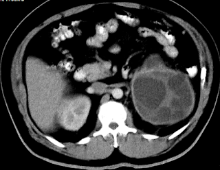
Here is a case of Renal Hydatid for the Radiology Grand Rounds submitted by Dr MGK Murthy and Dr Sumer Sethi of Teleradiology Providers. Concept and Archive of the Radiology Grand Rounds is available at- Radiology Grand Rounds


Echinococcosis is a worldwide zoonosis produced by the larval stage of the Echinococcus tapeworm. Adult worm lives in the proximal small bowel of the definitive host, attached by hooklets to the mucosa. Eggs are released into the host's intestine and excreted in the feces. Humans may become intermediate hosts through contact with a definitive host (usually a domesticated dog) or ingestion of contaminated water or vegetables. The ovum loses its protective layer as it is digested in the duodenum. Once the parasitic embryo passes through the intestinal wall to reach the portal venous system or lymphatic system, the liver acts as the first line of defense and is therefore the most frequently involved organ. Renal hydatid is rare accounting for 2% usually. There are no clincal symptoms except cystic rupture into the collecting system, which leads to acute renal colic and hydatiduria .

Imaging findings in hydatid disease depend on the stage of cyst growth (ie, whether the cyst is unilocular, contains daughter cysts, or is partially or completely calcified [dead]) . A difference in attenuation and signal intensity between the fluid in the central portion of the cyst and that in the peripheral cysts is a typical finding in echinococcosis due to a difference in content .Daughter vesicles (brood capsules) are small spheres that are formed from rests of the germinal layer and appear as cysts within a cyst. They contain the scolices and hooklets, along with sodium chloride, proteins, glucose, ions, lipids, and polysaccharides . When daughter cysts are separated by the hydatid matrix, they demonstrate a "wheel spoke" pattern .


Dr.Sumer K Sethi, MD
Consultant Radiologist ,VIMHANS and CEO-Teleradiology Providers

Sunday, June 29, 2008

Memories of a Nocturnal Walk in Boac (2)


It was the last night of my week-long sojourn to Marinduque. At eight in the evening I went out of my hotel room to gallivant once more the lovely little streets of Boac.

The evening was peaceful and cool with a lovely soft breeze of wind in the air, and it well reminded me I was one of the most rural places in the Philippines. The sky was well illuminated with the romantic glow of the full moon from the sky, creating lovely silhouettes of the ancient houses from beyond, dwarfed by the lovely clouds from above. On the small boulevards can be seen silhouettes of romantic lovers walking into the nearby town fair. Here and there people go to and from the fair.

The town was in a festive mood, the morrow being the the Grand Fiesta, the last day of celebrations. Overall, there was an ambiance of gaiety, and everyone expected it. The small bars were open up to the little wee hours of the morning offering the sleepless enjoyment and reverie. I stayed in one of the bars to drink a little and enjoy the sceneries and the playing live music.

It was past midnight when I started to walk back to my hotel room, still photographing street scenes. It was a lovely night, a memorable night.


Rizal Monument in the Town Hall


The Town Museum

One of the several old houses in Boac


The town fair


A street scenery


One of the few hotels in Boac


The old and dilapidated Dela Santa house, one of the oldest houses in Boac


The town market: closing time


A walk to remember

Saturday, June 28, 2008

The Risen Christ by Anastacio Caedo

The Risen Christ is a long lost work of the late noted sculptor Anastacio Caedo (1907-1990), protege and long-time assistant of Guillermo Tolentino. This sculpture was awarded the Gold Medal in the Art Exposition (sculpture category) of 1966 held at The Manila Hotel.

The Risen Christ is made of marble dust and stands at 1 and 1/2 feet tall. I think that it is one of the most beautiful Christ sculptures I have ever seen. I do not always collect sculptures, but when Caedo's heirs finally decided to let it have a new home, I knew exactly that it would be mine. It had brought great blessings in my life ever since.

Professor Anastacio Caedo is also known as the sculptor of several important historical monuments around the world, a few of which are the MacArthur Landing site in Leyte, the Mabini monument in Kalaw, the Ninoy Aquino monument in Makati, the Death March Memorial in Capas Tarlac, the Rizal monuments in Heidelberg and Wilhelmsfeld Germany, and the Juan Luna monument in Madrid, Spain.


Friday, June 27, 2008

Submandibular sialadenitis


Findings

There is diffuse soft tissue and glandular edema in the left sublingual and submandibular region. There is a large calcification in the left sublingual region representing a sialolith.

Differential diagnosis:
- Submandibular sialadenitis
- Submandibular carcinoma
- Malignant lymph node


Diagnosis: Submandibular sialadenitis


Key points

Presentation
- Unilateral painful submandibular gland (SMG) swelling with eating or salivation
- Painless mass

Treatment: Removal of submandibular gland in some cases.
Radiologic Evaluation: CT with contrast.

A sialolith is a calculus found in the salivary duct
- 85% are found in the submandibular duct, Wharton's duct
- 10% are found in the parotid duct
- 5% in the sublingual duct

Acute sialadenitis:
- Unilateral enhancing enlarged SMG with dilated duct behind the calculus
- Intraglandular ductal dilatation
- SMG cellulitis and/or myositis

Chronic sialadenitis:
- SMG small
- Fatty infiltration
- Intraductal calculus

Thursday, June 26, 2008

Cystic hygroma (lymphangioma)


Findings

Fetal MRI reveals a large mass extending from the anterior palate to the right supraclavicular region. It involves the anterior right neck of the fetus, as well as the right masticator space, parapharyngeal space, carotid, and parotid spaces. The airway is not well visualized, presumably due to airway compression.


Diagnosis: Cystic hygroma (lymphangioma)


Key points

Incidence is 1:5000.
Caused by absent or abnormal connections between the lymphatic and venous systems, or from sequestration of embryonic lymphatic tissue.
50% association with chromosomal abnormalities.
Often missed in the first trimester, as the most common initial finding of increased nuchal lucency is difficult to detect.
In the second trimester, is most commonly identified on ultrasound as a cystic mass with multiple thin walled septations, frequently with a posterior midline band, representing the nuchal ligament.
Important to identify anterior cervical soft tissue involvement because of increased morbidity from respiratory compromise.
Important to differentiate from posterior encephalocele, which is associated with an underlying skull defect, and cervical myelomeningocele, which is associated with an underlying vertebral defect.
MRI is usually not necessary in diagnosis, although it is helpful for surgical planning. In this case, Cesarean section with head delivery was performed at 40 weeks gestation to maintain uteroplacental circulation while tracheostomy was created because of anterior neck involvement and airway compression. Bulk resection was performed 3 weeks later.

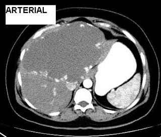
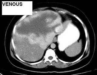
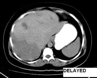
Giant Hemangioma of Liver-Triple Phase CT


Cavernous hemangioma is the most common benign hepatic tumor. It typically occurs in women. Lesions measuring more than 4 cm in diameter are known as "giant hemangiomas" and often cause symptoms such as vague abdominal distention and pain. The constellation of giant hemangioma, thrombocytopenia, and localized consumption coagulopathy is known as the Kasabach–Merritt syndrome. This is a case of 40 year old female with giant hemangioma of liver with triple phase CT showing classical centripetal fill-in.

Dr Jaya Shankar, MD and Dr.Sumer K Sethi, MD
Sr Consultant Radiologist ,VIMHANS and CEO-Teleradiology Providers

Monday, June 23, 2008

Silent sinus syndrome


Findings

Two coronal CT images (Figure 1 and Figure 2) and single axial CT image (Figure 3 ) demonstrate right maxillary sinus volume loss when compared to the left. There is inward retraction of the sinus walls (Figure 1, Figure 2, and Figure 3), increase in the size of the middle meatus (Figure 1 and Figure 2) and expansion of the retroantral fat (Figure 3). There is lateralization of the uncinate process and middle turbinate. This patient had concomitant right ethmoid air cell and frontal sinus opacification.


Diagnosis: Silent sinus syndrome


Silent sinus syndrome, or maxillary sinus atelectasis, is characterized by volume loss of the maxillary sinus after infundibular occlusion. It most often presents in the third through fifth decades of life with painless enophthalmos, facial asymmetry, and/or diplopia. Rarely do patients complain of symptoms of sinusitis.

Imaging findings of silent sinus syndrome are characteristic. There is maxillary sinus volume loss with inward retraction of the sinus walls and sinus opacification. The ethmoidal infundibulum is occluded, usually due to opposition of the uncinate process against the inferomedial orbit. The sinus volume loss accounts for the corresponding increase in ipsilateral orbital volume and size of the middle meatus.

The pathophysiology remains unclear. It is felt to be an acquired condition caused by chronic maxillary sinus obstruction and hypoventilation leading to negative intrasinus pressures. Chronic inflammation leads to osteolysis and thinning of the sinus walls which are retracted by the negative sinus pressure.

Treatment is aimed at creating an outlet for obstructed mucous via a nasal antral window or maxillary antrostomy. The goal of surgery is to prevent disease progression and further deformity.

Sunday, June 22, 2008

Hydatid Cyst- CT


Imaging--Separation of the laminated membrane from the penicyst produces a split wall or floating membrane appearance.

Dr.Sumer K Sethi, MD
Sr Consultant Radiologist ,VIMHANS and CEO-Teleradiology Providers
Editor-in-chief, The Internet Journal of Radiology
Director, DAMS (Delhi Academy of Medical Sciences

Stormy Weather

I went home early on Saturday because a strong typhoon is coming. "Frank", as the tropical storm is called, will be arriving in about the early morning hours of Sunday. It is Signal No.3, which means that it will hit Metro Manila directly.


Cubao rooftops on a stormy weather, the afternoon before typhoon Frank's arrival. View from my office window.

Prior to going home, I hoarded Farmer's grocery for some candles, mineral water, matchboxes, and batteries. Typhoons can cause power failures and it will be hours before electricity is restored. I saw that many people are doing the same in the grocery.

Outside, the gushing winds and heavy rains continue to flood the streets. Some street boys are running around enjoying the free shower, splashing themselves in the river of gushing waters. The scenery brought back distant memories of myself as a child. As a boy, I also begged my parents to let me shower in the rain. Sometimes, when my mother was in a good mood, I would be permitted. But there were times when I was not permitted (especially if I already took a bath before the rain began). Then, I would just look from our window, watching my friends in the rain as they play "habulang taga".

Back to the present day, going home was a trouble. The Jeepneys are nowhere to be found. Apparently, most of them went to garage early, expecting floods and heavy traffic. They weren't mistaken. In Aurora Boulevard, throngs of people filled the flooded streets waiting for taxis, FXs, jeepneys, or buses to deliver them home. It seemed that the streets will be filled with pedestrians rather than vehicles. From time to time, a few jeepneys and Fx passed by but already filled with passengers. The buses, when they come makes some big splashes of water that drenched the waiting pedestrians. One or two passengers debarked the bus, but ten or more replaced them so that the bus was filled to the entrance with people anxious to go home.

I decided to just walk the short distance from Aurora Boulevard to my house in New York, Cubao. It was now raining hard and I felt the cold wind in my face. My shoes, socks and my lower denims were already soaked.

Arriving home cold and wet, I decided to take shower. I boiled some water and poured it in my bucket. I also put some cold water, and after feeling that it was lukewarm enough, I enjoyed the soap and the bath. Feeling refreshed, I dressed in a fresh new shirt and shorts. Watched TV. There were 99 channels. All were boring. I decided to eat. And the best thing to eat in this cold inclement weather was hot instant cup noodles. I bought Nissin noodles from our local Sari-sari store. I love cup noodles but I hate the three minutes wait to cook it.

Outside, the gushing winds and rains continued. I was perfectly enjoying my noodles, even dipping my biscotcho in its steaming broth, when suddenly the power went out. I didn't expect that blackout will be this early. I lighted some candles and continued with my dinner. Afterward, I sat in my easy chair, and opened my laptop. Good thing that I charged it prior to the blackout. I decided to surf for some news on the net about the typhoon.

I felt bored and decided to play some pirated movies in my DVD collection. It was an old Italian movie called "The Bicycle Thief", a story about a man's desperate attempt to retrieve his stolen bicycle, because it's all the thing that he needs to be employed. Actually, I already watched it a few times before, but I love the simplicity of the story. I love movies with a touch of social realism. This preference of course saved me from watching again such trash as Indiana Jones and the Crystal Skull.

After a few hours, my laptop's batteries were drained. I fell asleep on my easy chair, only to be awakened at 3 a.m. by the howling winds and stronger rains. It was very dark as my lighted candles were already almost consumed. I looked outside and I saw that even the large tress in the neighborhood were swaying from the wrath of typhoon "Frank". Some tin roofs threatened to be blown away by the gushing winds. Once I felt safe that my own roof wouldn't give way, I slumped back into my easy chair.

I didn't have anything to do on a blackout and stormy night. So I slept again, while typhoon Frank made its way above my house and into the nearby provinces.

Saturday, June 21, 2008

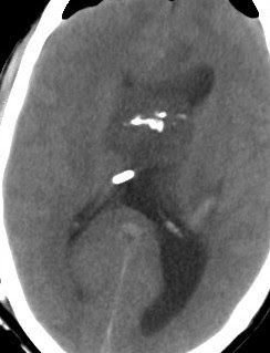
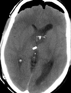
Central Neurocytoma-CT Imaging


Imaging of central neurocytoma is usually characteristic. Most of them occur as an exophytic, well circumscribed, globular mass that protrudes into the ventricles. Calcifications are common. This is a postoperative post shunting case of neurocytomas, note the intraventricular nature of tumour, calcification and operative pneumocephalus. Tumour showed immunoactivity for synaptophysin. Radiological differential diagnoses include oligodendroglioma, ependymoma, subependymal giant cell astrocytoma, and intraventricular meningioma.

Dr.Sumer K Sethi, MD
Consultant Radiologist ,VIMHANS and CEO-Teleradiology Providers

Thursday, June 19, 2008

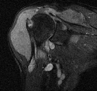
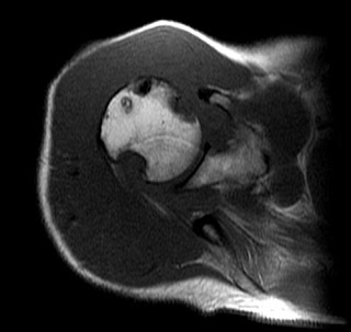
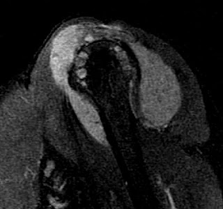
Pigmented Villonodular Synovitis of Shoulder


These are MRI pictures of a case of PVNS. Pigmented villonodular synovitis is well known in knee and shoulder involvement is reported rarely. Note the erosive defects in the humeral head.

Case by Dr MGK Murthy, Sr Consultant Radiologist
&
Dr.Sumer K Sethi, MD
Sr Consultant Radiologist ,VIMHANS and CEO-Teleradiology Providers

Contrast Induced Nephropathy

"In Radiology 2008;248:97-105 Shaun A. Nguyen et al compared effects of iso-osmolality contrast with a low-osmolality agent on renal function and concluded Intravenous contrast material application in high-risk patients is unlikely to be associated with permanent adverse outcomes. SCr levels after contrast material administration are lower in iodixanol than iopromide groups."

Wednesday, June 18, 2008

Sonographic gel put to novel use

Seung Ho Kim et al used sonography transmission gel as Endorectal Contrast Agent for Tumor Visualization in Rectal Cancer in MRI and concluded that it is an effective and safe endorectal contrast agent for rectal MRI. Published in AJR 2008; 191:186-189

Oral cavity dermoid


Findings

Axial contrast enhanced CT (Figure 1 and Figure 2) demonstrate a well circumscribed cystic mass (Figure 1) in the root of tongue. The lesion is heterogeneous with focal oval areas of fat attenuation within the lesion. There is a thin wall with no significant surrounding inflammatory changes (Figure 3). The lesion is in the midline between the genioglossus muscles (Figure 4).


Diagnosis: Oral cavity dermoid and epidermoid


Epidermoid and dermoid cysts are benign lesions encountered throughout the body, with 7% occurring in the head and neck area. The orbit is the most common site in the head and neck for these congenital lesions. They rarely occur within the oral cavity, representing less than 0.01% of all oral cavity cysts.

These congenital cysts are dysembryogenetic lesions that arise from ectodermal elements entrapped during the midline fusion of the first and second branchial arches between the third and fourth weeks of intrauterine life. Acquired cysts may be derived from traumatic or iatrogenic inclusion of epithelial cells or from the occlusion of a sebaceous gland duct.

The cysts can be classified as epidermoid when the lining presents only epithelium, dermoid cysts when dermal appendages are found, and teratoid cysts when other tissue such as muscle, cartilage, and bone are present. The teratoid type is the only variety that may have a malignant change.

Anatomically, these oral cavity cystic lesions most commonly involve the floor of mouth and may occur in the root of tongue (ROT), submandibular space (SMS) or sublingual space (SLS). Dermoid cysts generally present with slow and progressive growth, and even if they are congenital, the diagnosis is usually possible in the second or third decade of life. Midline cysts of the floor of the mouth present as painless subcutaneous or submucosal lesions. When large, they can displace the tongue and result in dysphagia, dysphonia or dyspnea.

Epidermoids present on imaging as low density, unilocular, well circumscribed simple cystic lesion. Dermoid cysts are usually more heterogeneous with fatty internal material and possibly calcification.

The treatment of dermoid cysts of the floor of the mouth is extracapsular excision with an intraoral or external approach, depending on the size of the lesion and the position relative to the mylohyoid muscle. The entire cyst must be removed to prevent recurrence.

Normal variant consistent with parietal foramina


Findings

On the ultrasound, there were 2 discrete bony defects in the skull, seen in the parietal regions bilaterally, that demonstrate sharp margins. These were each approximately 2 cm in diameter. The x-rays also demonstrate these same 2 round lesions.


Diagnosis: Normal variant consistent with parietal foramina


Key points

Parietal foramina are normal variants.
Normal parietal foramina transmit the emissary veins of Santorini (there can be a depression in the outer table at the vein's exit).
Defects are insignificant except in the differential diagnosis of bony defects (from surgical intervention or trauma).
These congenital defects have a characteristic location, but may vary in size.
These are often symmetric, although they can be asymmetric and irregular.

Tethered Spinal Cord


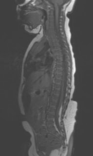
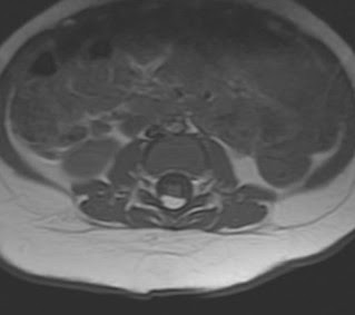
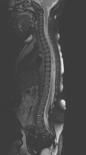
Here is a case of epidural/intradural lipoma showing signal suprresion on fat sat images and a low lying tethered spinal cord.

Dr.Sumer K Sethi, MD

Consultant Radiologist ,VIMHANS and CEO-Teleradiology Providers

Editor-in-chief, The Internet Journal of Radiology

Director, DAMS (Delhi Academy of Medical Sciences

Cord Astrocytoma


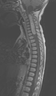
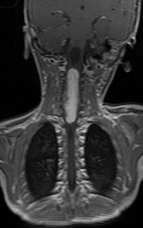
This is cervical cord astrocytoma with associated syrinx seen on Gd-MRI scan of a child.

Dr.Sumer K Sethi, MD
Consultant Radiologist ,VIMHANS and CEO-Teleradiology Providers
Editor-in-chief, The Internet Journal of Radiology
Director, DAMS (Delhi Academy of Medical Sciences