Sunday, May 31, 2009

Pancho Villa: First Filipino World Boxing Champion


The great symbol of the 1920s era in the Philippines was Pancho Villa, the most brilliant fighter of the period that bred such great boxers as Cabanela, Young Dencio, Frisco Concepcion, Clever Sencio, and the Flores Brothers.

Pancho Villa placed the Philippines on the map by winning boxing laurels abroad, defeating even the toughest flyweights in the United States. His fighting style was characterized by a relentless attack, a raging bull onslaught, and explosive and devastating punches.

His total fights of 105 (some only weeks in between) was a record in itself, elevating him into one of the great fighters in the history of boxing, and certainly one of the greatest Asian brawlers to step on the ring. The prestigious Ring Magazine, the bible of boxing aficionados, ranked Villa as one of the 100 Greatest Boxers of All Time.

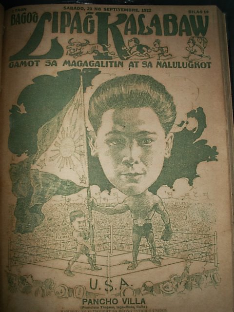
Cover for September 1922 issue of Lipang Kalabaw magazine.
Caricature by Fernando Amorsolo.Dennis Villegas collection.


Born Francisco Guilledo in Negros Occidental on August 1, 1901, he adopted the name Pancho Villa from the name of Mexico's famous revolutionary. Villa fought exclusively in the Philippines from 1919 through April 1922, often facing much larger men. In that period of time, he lost only three fights and captured two Filipino titles. In 1922, the American boxing promoter Frank Churchill discovered Villa in one of the amateur fights in Manila. Impressed by the young man's power punches, Churchill took Villa to the United States. The young Filipino fought two no-decision bouts in New Jersey, losing-according to the newspapers, to Abe Goldstein and Frankie Genaro.

The American press and public were at first slow to take notice of Villa. Churchill had difficulty arranging fights in major venues until, for almost no money, he got Villa and another Filipino, Elino Flores, on a card at Ebbets Field, home of the Brooklyn Dodgers. Each fighter won his bout, and the crowd gave Villa a standing ovation.

Cover for a September 1922 issue of Telembang magazine.
Portrait by Fernando Amorsolo.Dennis Villegas magazine collection

Three months after his arrival in the U.S., Villa knocked out American Flyweight champion Johnny Buff in the eleventh round to win the American flyweight title. To catch a glimpse of Villa's devastating attack, here's a very rare footage from his magnificent fight with Buff:


Genaro took the title back in 1923 in a 15-round decision that most observers believed belonged to Villa. Meanwhile, British flyweight champion Jimmy Wilde had come to New York seeking the world title. Wilde was then considered the best flyweight in the world. Although Genaro was a likely opponent, the now wildly popular Villa was considered a better draw.

In the much-anticipated match at New York's Polo Grounds on June 18, 1923, in front of thousands of spectators, Villa and Wilde set out for one of the most exciting fights in boxing history. Villa started slow, while Wilde started fast, throwing power punches that meant to knock-out the Filipino slugger. Villa defended successfully and threw some power punches of his own in retaliation, most of them landing and almost knocked down Wilde. In the second round and onwards, however,Villa started to display his relentless attacking style, peppering Wilde with punches from both hands. In the seventh round, Villa battered Wilde to a state of helplessness, knocking him
flat, face down in canvas, ending the fight --and Wilde's career. The crowd was ecstatic with Villa's victory--shouting "Viva Villa!" "Viva Villa!"

Here's a very rare footage of that famous bout, now considered one of the greatest slug fests in boxing history:


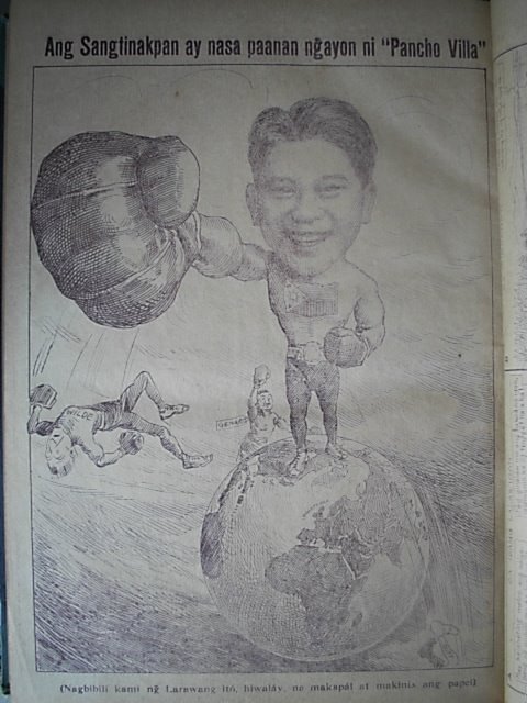
Pancho Villa caricature by cartoonist Jorge Pineda, Lipang Kalabaw 1923.
Dennis Villegas magazine collection

Villa was known during his time as being one of the cleanest boxers, always showing concern for his opponents and immediately turning away and walking to neutral corner after knocking down his opponent. This was before there was a rule of going to a neutral corner while the downed opponent is being counted by the referee.

Villa returned to the Philippines in September 1924, amidst jubilant reception (of his countrymen, not unlike the ones we do when Manny Pacquiao returns from a successful fight). He was invited for a parade and reception at the Malacanang Palace by then Governor General Leonard Wood, together with some of the big names in Philippine politics--then Senate President Manuel Quezon and House Speaker Sergio Osmena. It was known that General Wood and Senator Quezon were not in good terms, but the presence of the world champion temporarily set aside their personal differences.

As World Champion, Villa collected into his person all the swank and swagger of the era and the whole country felt an electrifying pride in his rise from rags to riches, his fetish for the most magnificent wardrobe, his expensive silk shirts and fashionable hats, his pearl buttons and gold cuff links, and his regal servants. He had a servant to massage him, another to towel him, a valet to put on his shoes, another to help him put on his trousers, still another valet to comb his hair, to powder his cheeks, and spray him with the most expensive perfume.

The Filipinos adored his extravagance, treating him almost as their crowned king. For a time, Villa was the most beloved figure in the Philippines--he had captured the heart and admiration of his countrymen, and he well thought he deserved it. He was perhaps more idolized as a showman, than as a boxer, and he was conscious of it. Never before had the Filipinos been electrified by the pride that their own kind had become the Champion of the World.

Villa successfully defended his title several times in the U.S. and the Philippines, and for a time, was considered practically invincible in the ring. Before returning to the United States, Villa defeated in Manila another great Filipino boxer, the mighty Clever Sencio. It was destined to be Villa's final victory in the ring--and no one among the thousands of cheering spectators knew it at that time.

In 1925, Villa fought in a non-title bout with Jimmy McLarnin in Oakland, United States. Weak from the recent extraction of a wisdom tooth, Villa lost the decision. It was destined to be his last fight. Another visit to the dentist resulted in the discovery of an infection and the extraction of three more teeth. Villa ignored the dentist's instructions to rest and return for a follow-up visit, and instead indulged in a week-long party.

The infection worsened, and by the time Villa's trainer, Whitey Ekwert, discovered the fighter's distress and rushed him to the hospital, it was too late. Villa died on July 14, 1925, of Ludwig's Angina, an infection of the throat cavity. He was survived by his wife Gliceria*.


Villa's untimely death at the young age of 24 broke the nation's heart. The hysteria that possessed the masses during his funeral was the most feverish of its era. Filipinos openly wailed in the streets while their hero's casket was being borne to its sad destination.

Such was the brief but shining career of one of the greatest Filipino boxers who ever lived.

panchovilla
Pancho Villa's grave inside the Manila North Cemetery.
The grave is being cleaned everyday by a tomb caretaker.

In 1989, Pancho's widow Gliceria- then 84 - insisted that a gambling syndicate conspired to murder the champion because of big losses in the Villa-McLarnin non-title fight. Pancho was a heavy favorite to beat McLarnin and the syndicate placed huge amount of bet to Villa. Mrs. Guilledo claimed that her husband was injected an overdose of anesthetic on instructions of the syndicate*.

In 1994, Villa was inducted posthumously in the International Boxing Hall of Fame, the second Filipino to earn the recognition--after Gabriel "Flash" Elorde.

*NY Times July 15, 1925: Villa "...died at a hospital here [San Francisco] today while undergoing an operation for an infection of the throat that developed from an infected tooth. Dr. C.E. Hoffman said the boxer suffocated under the anesthetic. Dr. Hoffman was preparing to operate when Villa's heart stopped. Artificial respiration failed to revive the patient."

Posterior Cruciate Ligament Avulsion Injury-MRI


Note the avulsion injury at the region of insertion of PCL.
Teleradiology Providers

Friday, May 29, 2009

Can You Guess This TV Theme? #12 - Answer

Awwwww Yeah! It's 'Family Matters' bitches! The spin-off from the show 'Perfect Strangers'.


The Winslow family, and their nerdy neighbor Steve Urkel, battled it out on my television set every Friday in my house.

Did you know that Reginald Vel Johnson (Carl Winslow) is the only actor to have appeared in all 215 episodes of the show? Kellie Williams (Laura Winslow) was in 214 episodes, and Jaleel White (Steve Urkel) was in 204 episodes.

The Winslows had a third child for the first four seasons. She was written out of the fifth season and all other future seasons and never mentioned again.... From the fifth season on, only Laura and Eddie Winslow were referenced....

Also, did you know that JudyAnn Elder replaced Jo Marie Payton as Harriet Winslow in the last half of the final season?

MVPuppets 3

NBA executives are praying to the basketball gods for a Los Angeles Lakers and Cleveland Cavaliers match-up in the NBA Finals. The most marketable thing ever on this planet is a Kobe Bryant v. LeBron James finals.

Anyway, I just found another one of those Kobe and LeBron commercials. It's super short, watch it....

Can You Guess This TV Theme? #12

I wanted to end the first dozen with a bang. Everyone can name this TV show theme.... I watch too much television!


Thursday, May 28, 2009

Retinoblastoma-CT


Note the soft tissue lesion with calcification in the left globe.

Can You Guess This TV Theme? #11 - Answer

Did you get it? Did you know the theme from one of the best television shows ever on television?


Yes, you did? Then it's time for the 'Dance of Joy'!


No, you didn't? 'Well, feed me garlic and call me stinky!'

Perfect Strangers was that show where a feet-on-the-ground guy from Wisconsin moves to the big city, and ends up taking in his nonsensical cousin who is fresh off the boat from Mypos.

Wait! Don't jump to conclusions! This show was not a one sided coin! Cousin Larry and Balki Batokomous were both able to teach each other a little bit about life throughout the show's 8 season run.

Did you know that Louie Anderson was cast for the role of Larry Appleton in the original pilot episode? Here's a still photo from the pilot. Would the show have been the success it was if Bronson Pinchot and Mark Linn-Baker weren't the stars?

Can You Guess This TV Theme? #11

I'm back from the face of the sun, I mean satan's taint, I mean the City of Lamont.

My dad was on this business trip with me, and stayed in the room next to mine. He said that when he pulled the comforter off of the bed, as soon as it hit the floor, he saw bugs scatter everywhere.

On a lighter note, here's today's theme.

Wednesday, May 27, 2009

Screw Lamont!

All right. I have one thing to say. Screw the City of Lamont! It smells like sulfur, and it's 95 degrees at 10:00 pm.

I'm staying at a creepy motel, called the Budget Inn. There are cigarette ashes all over the room, I'm afraid to take my socks off, and I'm not even able to enjoy the free Playboy Channel because I found what I can only assume is a pubic hair on what were supposed to be my clean sheets.

So, I'm writing this blog, while sitting in a chair near the room's window, wondering if I'm going to enjoy breakfast at 'Le McDonald's' or 'Le Jack in The Box'. Both of which were recommended to me when I asked the manager of the hotel, "Where is a nice place to eat breakfast around here?"

I'm not going to post "Can You Guess This TV Theme" today. I'm too consumed by the pair of eyes, I can only hope I am imagining, are staring at the me through the motel's bathroom windows. Why are they clear? Aren't the windows in bathrooms supposed to be frosted?

So, I'm going to post a video.... If you were a geek at any point during the previous 35 years, you should be able to relate to this video.... Laugh at me, I dare you, I'll throw my twelve-sided die and attack you with my goblin sword....


Ocular Wegener’s granulomatosis


Findings


There is an extraconal mass within the right medial orbit, adjacent to the lamina papyracea, extending in an arc like fashion from the superomedial aspect to the inferolateral aspect of the right orbit. Mass extends into the intraconal space and splays the medial and inferior rectus muscles. Mass is hypointense to cortex on T2 weighted images and isointense to cortex on T1-weighted images. Mass demonstrates homogeneous post contrast enhancement. There is associated proptosis of the right globe. There are extensive postoperative changes of the paranasal sinuses and mucosal thickening involving all of the paranasal sinuses, predominately the ethmoid air cells.

Differential diagnosis:
- Sarcoidosis
- Lymphoma
- Vasculitis / angiitis (e.g., Wegener's granulomatosis)
- Subperiosteal abscess


Diagnosis: Ocular Wegener’s granulomatosis (favored diagnosis: the patient was known for Wegener’s granulomatosis)


Discussion

Wegener's granulomatosis is a multisystem disease classically characterized by necrotizing granulomatous inflammation of the upper and lower respiratory tract and the kidneys and necrotizing vasculitis of the small and medium sized vessels. Patients typically present between 35-55 years of age. The nose and sinuses are most commonly affected, and patients may present with sinus headaches and nasal drainage. The lungs are affected in 85% of patients, and kidneys are involved in 75% patients. Most of the morbidity of the disease is usually related to the renal component.

Wegener's involvement of the eye is not uncommon, occurring in 30-60% of cases. Approximately 8-16% of patients with Wegener's granulomatosis initially present with ophthalmic disease. Ocular manifestations can be diverse, ranging from mild conjunctivitis to episcleritis, uveitis, ciliary vessel vasculitis, and retro-orbital mass lesions. Vision loss can occur secondary to mass effect, ischemia, or vasculitis of the optic nerve or occlusion of the retinal artery.

Several medication regimens utilizing immunosuppressants are given to patients with Wegener's granulomatosis. Commonly used drugs include steroids, cyclophosphamide, azathioprine, and methotrexate.


Key radiology findings in Wegener's granulomatosis (head and neck)

Paranasal sinus inflammatory disease associated with nodular soft tissue mass in the nasal cavity.
Destructive/erosion changes of the nasal septum; nasal septum perforation is common.
Orbital invasion is the most common site of paranasal sinus disease extension.
Orbital findings can vary but may include, retro-orbital soft tissue mass resulting in compression of adjacent structures. Although rare, orbital granulomata can calcify.

Tuesday, May 26, 2009

Can You Guess This TV Theme? #10 - Answer

Tom Hanks and Peter Scolari play Kip Wilson and Henry Desmond who play Buffy and Hildegarde in 'Bosom Buddies'.


The theme to the show was Billy Joel's "My Life". The show was really funny! Tom Hanks and Peter Scolari have great chemistry, however, they're not attractive women.

And how can you lose with Tom Hanks? The man can do drama, the man can do comedy... And he's one of the most loved and most successful men in Hollywood. He can't lose!

How do you think that pitch meeting went? 'Ok, I have an idea for a show.... There's these two guys, their apartment is destroyed... So they have to get another affordable apartment.... But the only way they can do that, get this, you're gonna laugh, they have to dress as women because it's an all female building. Wait.... It gets better! None of the women in the building will be able to figure out that the two characters are men in women's clothing. Just to put the icing on the cake... We have two unknown actors for the lead roles!'

Can You Guess This TV Theme? #10

This show only lasted two seasons, and if I give any more hints I'll give it away. So here it is, give it your best shot....

Monday, May 25, 2009

Can You Guess This TV Theme? #9 - Answer

I was going to add some clever comment in the blog this morning... Something like 'You should be able to identify this easily, however, I'd identify it using a paperclip, a wad of chewing gum, and spectacles from the 1940's'.

I'm sure you are all very sad that you missed out on this humor, however, as I'm writing this, I'm still giggling to myself. 'Spectacles.... That's an old-timey word for glasses.... Giggle Giggle'.

All right, so by now, I'm assuming that you all knew that the theme was from the television show "MacGyver".


Did you know that MacGyver's first name was Angus?
Richard Dean Anderson is awesome...

Can You Guess This TV Theme? #9

Being that it's Memorial Day, I wanted my 'Can You Guess This TV Theme' to have a military theme. Naturally, you would think MASH, right? Yeah, I tried that, however, the friendly people over at YouTube will steal your first born child if you try to upload any original MASH content. I couldn't find any good MASH videos, and I never understood why that show was so popular, so I have something better for you all.

So, here's today's TV theme. Enjoy!


Sunday, May 24, 2009

Spontaneous rupture of arachnoid cyst into subdural space


I was asked for opinion when i was just walking by the OPD of hospital, couldn't resist clicking the image with my mobile phone. Hence the poor quality of image but the finding is well seen. Arachnoid cysts are known to present as spontaneous subdural hygroma. Also noted a snap shot of my Teleradiology centre in Delhi.


Side Discrepancy Errors

"Side discrepancy errors in radiology reports do occur and it is important that radiologists, referring physicians and patients communicate well to help prevent errors in clinical management." Reports Medical news today

Saturday, May 23, 2009

MVPuppets 2

What? A Saturday post? Crazy right?

Anyway, I just found another one of those Kobe and LeBron commercials. Here it is for your viewing pleasure.

Diastematomyelia


Findings

Figure 1: Myelogram of the thoracolumbar spine demonstrates a vertical linear cleft within the distal cord dividing it into two hemicords. Vertebral and rib abnormalities are noted in the lower thoracic spine.
Figure 2: Axial post myelogram CT image of the thoracic spine shows two hemicords.
Figure 3: Axial T2-weighted MR of the thoracic spine shows two hemicords.


Diagnosis: Diastematomyelia


Diastematomyelia is characterized by a sagittal division of the spinal cord into two hemicords, each with one central canal, dorsal horn, and ventral horn. An osseous or fibrous septum separates the two hemicords. The condition represents a disorder of neural tube fusion with the persistence of mesodermal tissue from the primitive neurenteric canal acting as the septum or cleft. 85% of clefts occur in the thoracolumbar spine between T9 and S1. The hemicords typically reunite above and below the cleft.

Two types of diastematomyelia have been defined:
- Type 1 diastematomyelia: each hemicord has its own dural sheath
- Type 2 diastematomyelia: both hemicords are covered by a common dural sheath

Congenital spinal deformities are seen in approximately 85% of patients with diastematomyelia. Intersegmental laminar fusion is considered virtually pathognomonic for the condition. Scoliosis, tethered cord, spinal dysraphism, and syringohydromyelia are also commonly associated.

Diastematomyelia occurs in 5% of patients with congenital scoliosis. The diagnosis is typically made in childhood, and is much more common in females. The clinical presentation varies from asymptomatic to neurologic abnormalities indistinguishable from other causes of a tethered cord. Cutaneous stigmata, most commonly a “fawn’s tail” hair patch, may indicate the level of diastematomyelia. The treatment of symptomatic patients is excision of the bony spur or septum and lysis of the adjacent adhesions.

Cortical Hyperintensity on DWI


Diffusion-Weighted Imaging in the Setting of Diffuse Cortical Laminar Necrosis and Hypoxic-Ischemic Encephalopathy. Note the cortical hyperintensity on diffusion weighted imaging.

Friday, May 22, 2009

Symptomatic os subfibularis-MRI


Separated ossicles at the tip of the lateral malleolus, the condition known as os subfibulare, are sometimes a cause of ankle pain. There are two theories regarding the origin of os subfibulare. One theory proposes that it is caused by an avulsion fracture attributable to pull of the anterior talofibular ligament, whereas the other theory proposes that it is the result of an accessory ossification center. This is 24 year old male with chronic ankle pain.

Can You Guess This TV Theme? #8 - Answer

I just finished watching the entire series of "My So Called Life" on DVD.


Screw "My So Called Life"! How dare this show take my gentle soul and crush it into a million pieces. I am not happy! Are Angela Chase (played by Claire Danes) and Brian Krakow going to get together? Is she going to stay with Jordan Catalano (played by Jared Leto)? What's going to happen with the gay friend? Or the druggie friend? Or the goodie two-shoes friend? Will there be harmony ever again? It just ended with a season 1 cliffhanger! There are so many doors left open!!!!!!! I need to know what happens!!!!!!! Please god!!!!!!! I need to know what happens!!!!!!!

I've looked everywhere! Not one person gives any kind of closure as to what could have happened after the first season. And when I say I looked everywhere, I mean I looked at the first two sites that came up when I googled "My So Called Life Season 2".

By the way, Colleen, you're right. Jordan Catalano is dreamy!!!!

MVPuppets

A match up in the NBA Finals between two of the greatest players in the history of the sport (Kobe Bryant and LeBron James) seems to be inevitable.

Nike has put together a few videos with Kobe and LeBron puppets... They're hilarious! Here they are.... In the "Chalk" commercial, it looks like Kobe and LeBron live in a house filled with mountains of cocaine.


Minnie Lost Her Mickey

Wayne Allwine, the actor who voiced Mickey Mouse for the last 32 years, died on Monday, May 18, 2009, from complications of diabetes.

He is survived by Russi Taylor, his wife of 18 years, and the current voice of Minnie Mouse (and has been for the past 23 years).

Minnie lost her Mickey.... Awwwwwww.......

Can You Guess This TV Theme? #8

Here's an easy one.... Colleen, I'm going to throw you a bone this time. You should be able to answer this one immediately.... I proudly retain my status of having the same television tastes as a 13-year old girl....

Pneumocele of the sphenoid sinus with CSF fistula


Findings

Figure 1: Axial CT in bone windows demonstrates expansion of an air-filled right sphenoid sinus with thinned and eroded walls. Pressure erosions at the base of the right pterygoid process, medial wall of the right foramen ovale, and anterior surface of the clivus are evident.
Figure 2: Axial CT in bone windows again shows the expanded air-filled right sphenoid sinus. Note that the wall of the right carotid canal is dehisced, leaving the right internal carotid artery (ICA) completely exposed.
Figure 3: Axial CT in bone windows shows narrowing of the right superior orbital fissure by the expanded right sphenoid sinus. Again seen is complete exposure of the right ICA.
Figure 4: Reformatted coronal CT in bone windows demonstrates the expanded air-filled right sphenoid sinus eroding the base of the right pterygoid process and effacing the ipsilateral foramen rotundum.
Figure 5: Reformatted coronal CT in bone windows shows the expanded air-filled right sphenoid sinus eroding the base of the right pterygoid process and effacing the ipsilateral vidian canal.
Figure 6: Reformatted sagittal CT in bone windows shows the expanded air-filled right sphenoid sinus eroding the clivus and thinning the dorsum sellae.
Figure 7: Axial image from CT cisternogram demonstrates intrathecal contrast leaking into the expanded right sphenoid sinus along the septum.
Figure 8: Direct coronal image from CT cisternogram again shows intrathecal contrast leaking into the expanded right sphenoid sinus along the septum. (Note that the image is horizontally flipped).


Diagnosis: Pneumocele of the sphenoid sinus with CSF fistula


The term pneumocele refers to an aerated sinus that is enlarged beyond normal anatomic margins and whose walls demonstrate focal or diffuse wall thinning or erosion. Pneumosinus dilatans also describes abnormal sinus expansion but with normal wall thickness and integrity. Several past articles on this topic have used the term pneumosinus dilatans to describe an abnormally expanded sinus with or without bony erosion, favoring a single term to describe what the authors believed to be variations of the same entity. We prefer to distinguish between the two entities based on the presence or absence of bony wall thinning or erosion. Nevertheless, both of these lesions can occur in any sinus, but the maxillary sinus is most commonly involved by pneumoceles, whereas the frontal sinus is most commonly affected by pneumosinus dilatans. In contrast, pneumocele of the sphenoid sinus is a rare lesion.

Pneumoceles may occur suddenly, presumably by a trap-valve mechanism or by rupture of a large mucocele. In contrast, pneumosinus dilatans tend to develop chronically. However, the etiology and pathogenesis of pneumosinus dilatans are still poorly understood. Proposed theories of sinus hyperpneumatization include infection with gas-forming microorganisms, hormonal influences, and congenital defects. Pneumosinus dilatans are usually incidental findings, but have been associated with various disorders including but not limited to fibrous-osseous dysplasia, Klippel-Trenaunay, hydrocephalus and arachnoid cysts. Pneumosinus dilatans of the sphenoid sinus may also be the first sign of a meningioma of the planum sphenoidale or tuberculum sellae.

The clinical severity of symptoms depends upon the sinus or sinuses involved and will guide patient management. Reported manifestations of pneumoceles, and similarly of pneumosinus dilatans, include but are not limited to headaches, frontal bossing, exophthalmos, vision loss, and CSF rhinorrhea. Based on the presumed etiology of a trap-valve mechanism, the surgical treatment of pneumoceles includes opening the affected sinus directly into the nasal cavity via transnasal endoscopic approach. In our case of sphenoid pneumocele with associated CSF fistula, the sphenoid sinus was packed with subcutaneous abdominal fat using transnasal endoscopic technique.

Patellar Tendon Injury-MRI


Thursday, May 21, 2009

Pulley Lesion-Shoulder MRI


FINDINGS:
The alignment of the different structures of the shoulder joint are well preserved. There is no evidence of instability, there is no evidence of dislocation. The bone marrow signal of the different bony structures is normal. No abnormalities are identified to the bone marrow. There is no evidence of microtrabecular lesion, hemorrhage, bony bruise, cortical disruption or fracture. There is evidence of osteoarthritis identified to the glenohumeral joint, with evidence of altered signal intensity in the superior labrum in its posterior part consistent with a SLAP lesion. Also noted is fluid in the subcoracoid bursa in relation to the superior part of the subscapularis tendon insertion likely consistent with a rotator interval tear. Long head of biceps shows some evidence of altered morphology and medial subluxation. . At the glenoid surface, there is evidence of subchondral sclerosis. There is evidence of synovial fluid in the joint in relation to the labrum and glenoid surface, distending the subglenoid recess. Fluid is also noted in the subdeltoid and subacromional bursa. There is evidence of osteoarthritis identified to acromioclavicular joint with irregularity of the articular surface, capsular distension. Acromion process shows minimally curved undersurface. There is evidence of altered marrow signal intensity in relation to the greater tuberosity of humerus with increased marrow signal in relation to the insertion of the suprasinatus and infraspinatus muscles consistent with insertional enthesopathy. The tendons of rotator cuff including supraspinatus, infraspinatus, subscapularis and teres minor tendons are normal. The bone marrow signal of the rest of bony structures is normal. No abnormalities are identified to the insertions of the deltoid.

IMPRESSION:
1. The findings are consistent with osteoarthritis of the glenohumeral and acrominoclavicular joint.. At the level of the joint, there is fluid in relation to labrum and glenoid surface, distending the inferior recess, in subdeltoid and subacromional bursa
2. There are findings consistent with posterosuperior labral tear (SLAP lesion) and rotator interval tear with fluid in relation to the superior border of subscapularis & subcoracoid bursa. 3. There is some atrophy of the long head of biceps and medial subluxation (may be a result of the “pulley lesion”)
4. Insertional enthesopathy in the humeral head in relation to supraspinatus tendon
5. No evidence of tear or retraction of the supraspinatus tendon is identified.
6. No evidence of a microtrabecular lesion was identified.

Can You Guess This TV Theme? #7 - Answer

I remember waking up early every morning during summer vacation one year, just so I could watch "Mama's Family" in syndication.

Vicki Lawrence played Thelma "Mama" Harper, cranky queen of the family providing shelter and knocking sense into her dimwitted family. Her son Vinton and his wife Naomi, her nephew Buddy and her nosy neighbor Iola made up her gang of dolts.

I was watching clips of "Mama's Family" on the computer today.... Yeah.... I don't find the show quite as funny as I did 20 years ago....

Can You Guess This TV Theme? #7

Ok Jerry.... You've guessed them all so far.... Try this one.... This show was very popular, but the theme is just generic enough to make it sound like every other theme for every other television show ever....

HIV encephalitis


Findings

Mild parenchymal volume loss with symmetric nonenhancing periventricular and subcortical white matter T2 prolongation. No mass effect.

Differential diagnosis:
- Microvascular disease
- HIV encephalitis
- PML
- Toxo/lymphoma
- Chemotherapy/Radiation
- Leukodystrophy
- Demyelination – MS, ADEM


Diagnosis: HIV encephalitis (known HIV infection).


Key Points: HIV Encephalitis / AIDS Dementia complex

Cognitive disturbances that progress to dementia
7-27% of AIDS patients
Direct CNS infection by HIV +/- CMV
MR imaging primarily shows central volume loss and diffuse or periventricular white matter disease (both demyelination and gliosis)
Can be difficult to differentiate from PML
Pre-HAART (Highly Active Antiretroviral Therapy) prognosis was poor, but HAART therapy is felt to decrease incidence and induce remission.

Wednesday, May 20, 2009

Can You Guess This TV Theme? #6 - Answer

Jerry, you're too good.... You are correct, today's theme was the song "C'mon C'mon" by the Von Bondies, and it's the theme for the television show "Rescue Me".


Do any of you watch this show? It is fantastic! I just finished watching the fourth season....

The character development is amazing. After watching the first three episodes, you'll feel like you've known the characters your entire life, and you'll have to know what happens next. The show is emotional and passionate. It's just a fascinating hour of television.

Denis Leary plays an ill-tempered, self destructive, hypocritical, manipulative, relapsed alcoholic fireman who has to deal with his crazy family, crazy job, crazy coworkers and a crazy post 9/11 world. He plays the role sooooo well.

My grandfather was a New York fireman, and it's like I can see him in the characters.

You can watch all of the episodes at Hulu for free.... I'm so nice.... I'll help you get there.... Just click on the logo below....

Can You Guess This TV Theme? #6

This is the theme from a television show that is still on TV, however, nobody else I know watches it. So I'm going to guess that not one person who reads my blog will know what show this theme is from.....

Cerebral amyloid angiopathy (CAA)


Findings


MRI of the brain without contrast shows diffuse cortical atrophy as well as multiple small (< 5 mm) cortical-subcortical foci of decreased signal intensity, best seen in GRE sequences (Figure 7 and Figure 8). Some of these focal areas of low signal intensity are not seen or are barely discernible on T2-weighted sequences (Figure 3 and Figure 4), and not seen at all on T1-weighted (Figure 1 and Figure 2) or FLAIR sequences (Figure 5 and Figure 6). No areas of abnormal signal intensity are seen in the basal ganglia. In addition, areas of increased white matter signal intensity, compatible with leukoencephalopathy, are best seen on FLAIR sequences (Figure 5 and Figure 6).


Diagnosis: Cerebral amyloid angiopathy (CAA)


Cerebral amyloid angiopathy (CAA) is a significant cause of cortical-subcortical cerebral bleeds in the normotensive elderly individual. In CAA, beta-amyloid protein is deposited in the media and adventitia of small to medium sized blood vessels in a cortical, subcortical, and leptomeningeal distribution. These deposits are associated with fibrinoid necrosis, vessel wall fragmentation, and microaneurysms, all of which produce vascular fragility and lead to spontaneous micro and/or macrohemorrhages, with the latter sometimes having devastating consequences. Fibrinoid necrosis can also lead to blood vessel narrowing and subsequent distal ischemia. CAA is sometimes called Congophilic amyloid angiopathy, because beta-amyloid deposits are highlighted with Congo red stain and show yellow-green birefringence under polarized light.

CAA has been found at autopsy in up to one third of individuals between 60 and 70 years old, and in up to two thirds 90 years and older. Nevertheless, the majority of patients with CAA remain asymptomatic, and thus the condition is currently under recognized. When symptoms do arise, they are similar to those of a transient ischemic attack or dementia, which are nonspecific findings making CAA difficult to diagnose clinically. Patient symptomatology is usually related to macrohemorrhages, defined as larger then 5mm, and can resemble an acute ICH. CAA is not associated with systemic amyloidosis, yet is strongly associated with Alzheimer disease.

Radiographic findings of CAA normally include cortical and subcortical hemorrhages, atrophy, and leukoencephalopathy. Microhemorrhages associated with CAA are normally not seen on CT, T1-weighted, or T2-weighted sequences. Therefore, if CAA is suspected clinically, or in the event that cortical-subcortical focal intracranial hemorrhages are seen on a noncontrast enhanced CT, an MRI should be obtained, which includes T2*-weighted gradient-echo (GRE) sequences. At present, GRE is one of the most sensitive sequences for the detection of acute and chronic hemorrhages, like the ones associated with CAA. The hemosiderin present within these cerebral hemorrhages, causes local magnetic field inhomogeneities, with a prominent loss of signal on T2*-weighted GRE sequences. Susceptibility-weighted imaging (SWI) is another sequence that is gaining acceptance, which is also very sensitive in detecting cerebral microhemorrhages. Nevertheless, for a definitive diagnosis of CAA, biopsy or autopsy is required. It is important to note that patients taking anticoagulants or aspirin which are diagnosed with CAA, should be counseled regarding the risks and benefits of treatment. Finally, no treatment is currently available to halt or reverse beta-amyloid protein deposition associated with CAA.