Friday, July 30, 2010

Cerebello-pontine Angle Epidermoid-MRI

On T1-weighted images, epidermoids are generally slightly hyperintense or isointense relative to gray matter. The lesions are usually isointense relative to CSF on T2-weighted images, but they may be slightly hyperintense. The center of these lesions usually has an internal architecture with areas of heterogeneity. Enhancement of portions of the rim may be seen after the administration of contrast material. The remainder of the lesion does not enhance. Diffusion-weighted imaging can now be used for these lesions epidermoids have markedly restricted diffusion and, therefore, high signal intensity on the diffusion-weighted trace images.


Intramedullary spinal cord metastases


Findings

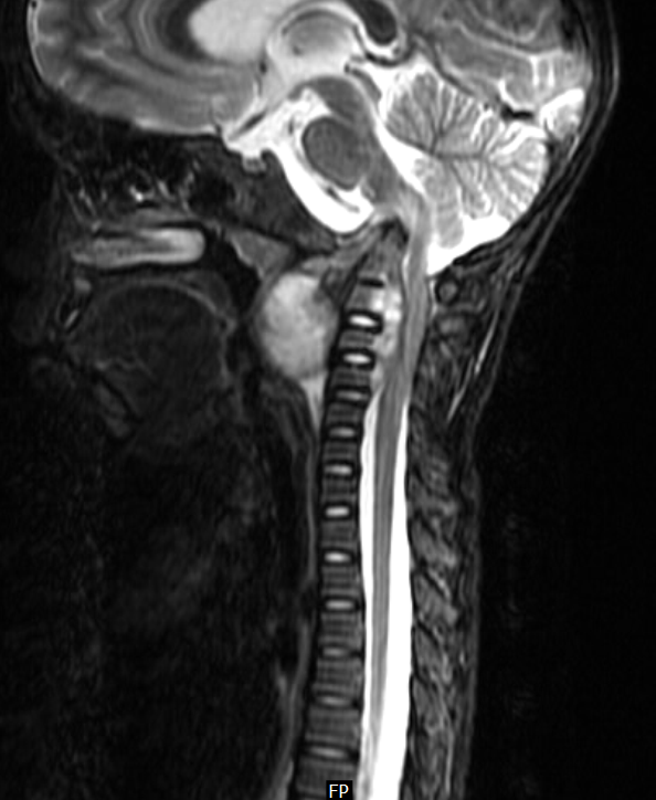
Figure 1: Sagittal T1 weighted image shows focal expansion of the cord substance in the region of the upper dorsal vertebrae (T3 and T4). The area is isointense to the cord.
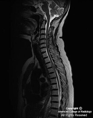
Figure 2 and Figure 3: Sagittal T2 weighted and STIR images show a hyperintense lesion involving the dorsal spinal cord with edema, seen at its rostral and caudal ends. Incidentally seen is a hemangioma (Figure 1 and Figure 2) involving the T7 vertebral body.

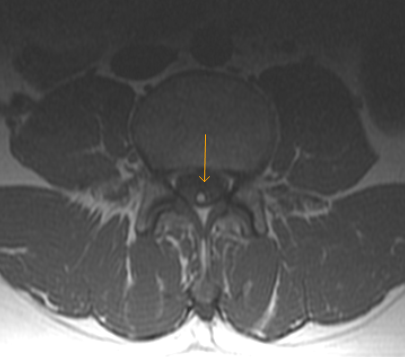
Figure 4 and Figure 5: Post contrast axial and sagittal T1 weighted images show almost homogeneous enhancement in the lesion. Incidentally seen are pleural effusion (Figure 5) and pulmonary metastasis (Figure 5).


Diagnosis: Intramedullary spinal cord metastases (ISCM)


The most common neurological complications of breast cancer are brain metastases and spinal cord compression. In most instances spinal cord compression is caused by extra-dural soft tissue masses, however ISCM is a rare and a distinct diagnostic possibility.

ISCM is an unusual presentation of systemic malignancies. Close to 50% of all ISCMs arise from primary lung tumors, with small cell carcinoma being the most common. The remainder originate from primary cancers of the breast, colon, melanoma, lymphoma, and kidney. ISCMs are typically solitary and extend over a length of 2-3 vertebral segments.

The clinical manifestations of metastatic intramedullary spinal cord tumors are typically back pain, paresthesia, paraparesis, spasticity of the lower extremities, and autonomic dysfunction.

Magnetic resonance imaging is considered the gold standard for the diagnosis of tumors affecting the spinal cord. The typical ISCM seen on MRI is a small, isolated, oval-shaped lesion with or without slight deformation of the spinal cord profile. It is isointense on T1-weighted images with a nodular contrast enhancement and a pencil-shaped hyperintensity on T2-weighted sequences, most frequently extending proximal to the lesion. Cysts are rare, in contrast to primary intramedullary neoplasm.

External beam radiation with or without concomitant corticosteroids has been the most effective method of treating ISCM. In a small group of selected patients, surgical resection seems be a reasonable option, especially in cases presenting with previously undiagnosed or limited primary tumors and rapid neurologic deterioration. Also, when the primary tumor is well known to be radioresistant, as in the case of melanoma or renal cell carcinoma, surgical decompression or subtotal resection would be indicated.

Patients with ISCM have a very short life expectancy; their median survival is 3 to 4 months from the time of diagnosis. Those with breast cancer as the primary source of ISCM, tend to do better than other types of cancer; their median survival is 13 months.

Venous Angioma-MRI

On contrast-enhanced MRI, the cluster of veins in developmental venous anomalies has a spoke-wheel appearance; the veins are small at the periphery and gradually enlarge as they approach a central draining vein. This appearance has been referred to as caput medusa, or the head of Medusa, because of the serpentine appearance of the curvilinear peripheral draining veins.  These are axial and coronal post contrast images of 13 year old girl with hsitory of absence seizures. Reported by- Teleradiology Providers


Thursday, July 29, 2010

CV Junction Potts Spine-MRI

Cervical Pott’s disease is unusual. Furthermore, CVJ tuberculosis has been reported to affect 0.3% to 1% of all Pott’s disease patients. This is a 9year old boy with potts spine with cranial migration of the odontoid process and retropharyngeal abscess. Note the impingement of brainstem with odontoid process.  The occipito-cervical junction, a transitional zone between the skull and the spinal column, serves as the most mobile part of the axial skeleton. Bony abnormalities affecting this complex results in dysfunction of the neural structures by compression along the entire circumference, altering the arterial supply, venous drainage and changing the CSF fluid dynamics.


Monday, July 26, 2010

Chronic progressive external ophthalmoplegia


Findings

The extraocular muscles are atrophic and to some degree show fatty replacement. No abnormal enhancement is present. The globes are intact bilaterally. The intraorbital fat appears slightly increased. There is bilateral orbital proptosis with both globes anterior to the interzygomatic line.


Diagnosis: Chronic progressive external ophthalmoplegia


Key points

Chronic progressive external ophthalmoplegia (CPEO) is characterized by slowly progressive paralysis of the extraocular muscles.
Mitochondrial myopathy usually associated with skeletal muscle weakness.
Presents with bilateral, symmetrical, progressive ptosis, followed by ophthalmoparesis months to years later.

Kearns-Sayre syndrome: Related mitochondrial myopathy with CPEO, onset before age 20 years, pigmentary retinopathy, and at least one of the following: cardiac conduction defects, CSF protein of greater than 100 mg/dL, and/or cerebellar syndrome.
KSS can include mental retardation, hearing loss, seizures, short stature, delayed puberty, and various endocrine disorders.
Frequency: Rare. Males=Females.

Imaging studies: MRI, CT, and ultrasound may show thin, symmetrical extraocular muscles in CPEO, in contrast to enlarged extraocular muscles sometimes seen with Graves disease.
Those with KSS and CPEO display a wide spectrum of MRI findings, including normal brain, diffuse atrophy, and T2 prolongation in subcortical cerebral white matter, cerebellar white matter, globi pallidi, thalami, and substantia nigra.
Diagnosis: Muscle biopsy is definitive test but PCR also shown to be conclusive.

Saturday, July 24, 2010

Lipomatous Filum terminale

Fat in the filum terminale is not an infrequent occurence, seen in 4 - 6% of individuals , and is especially easily detected on MRI. It is usually an incidental finding of no clinical concern.  There were no related symptoms and no evidence of tethering. The thickened fatty filum terminale seemed to be a developmental anomaly and without clinical significance.


Friday, July 23, 2010

Hairline fractures and Teleradiology

I am not sure if this has been reported before but problem with online reporting is not with complex disorders or with syndromes as our team compromises of senior rads and they are well versed with these situations. Problem possibly is with hairline fractures.

Usually we get no clinical information at all especially in orthopaedic plain films let alone any information on point of tenderness, so we are likely to miss some of the hairline injuries and as soon as we miss any findings we being a teleradiology company cases get bounced back to us very fast and referring physicians think we missed a simple HAIRLINE FRACTURE!

Do you also have similar experiences with reading plain orthropedic films without history?

Wednesday, July 21, 2010

Moya Moya Disease-MRI

Moya Moya is a rare idiopathic vasoocclusive disease characterized by progressive irreversible occlusion of main blood vessels to the brain as they enter into the skull. The occlusive process stimulates the development of an extensive network of enlarged basal, transcortical and transdural collateral vessels . In Japanese, Moya Moya means hazy. The disease derives its peculiar name from the angiographic appearance of cerebral vessels in the disease that resembles "puff of smoke". The process of blockage, once it begins tends to continue despite any known medical management unless treated with surgery.

MRI not only reveals areas of infarctions but also allows direct visualization of these collateral vessels as multiple small flow voids at the base of brain and basal ganglia. MR angiography is used to confirm the diagnosis and to see the anatomy of the vessels involved. It typically reveals the narrowing and occlusion of proximal cerebral vessels and extensive collateral flow through the perforating vessels demonstrating the classic puff of smoke appearance.

Our patient is 17 yr old female with history of recurrent seizures. MRI T1 weighted images show extensive collateralization of vessels in region of circle of willis. MRA reveals reduced luminal caliber of left internal carotid artery in petrous, cavernous and supraclinoid segments. Left MCA also shows attenuation of its luminal caliber when compared to its counterpart on right side along with collaterals at the base of the brain and basal gnaglia.


Second Opinion By -Teleradiology Providers

Friday, July 16, 2010

Mesial temporal sclerosis with infarct of the parahippocampal gyrus


Findings

There is expansion and abnormal FLAIR and T2 signal within the right hippocampal formation and parahippocampal gyrus. There is diffusion restriction of the parahippocampal gyrus.


Diagnosis: Mesial temporal sclerosis with infarct of the parahippocampal gyrus


Discussion

Mesial temporal sclerosis (MTS) is a poorly understood phenomenon involving atrophy and sclerosis of the hippocampus and adjacent structures, namely, the amygdala, parahippocampal gyrus, and uncus. MTS may be acquired in the setting of prolonged febrile seizures, status epilepticus, or cerebral ischemia. Histopathology demonstrates neuronal loss and fibrillary gliosis. There has been controversy regarding whether MTS is the cause or the result of temporal lobe epilepsy. However, there is a clear connection since the majority of temporal lobe resection specimens done for temporal lobe epilepsy demonstrate MTS. Approximately 15% of temporal lobe resection specimens exhibit both MTS and another lesion such as cortical dysplasia. Approximately 25% of patients are successful with medical therapy. The patient presented here had longstanding seizures and had a recent seizure which resulted in acute infarction of the parahippocampal gyrus. This may result from seizure related hypoxemia.


Radiologic overview

MR demonstrates increased T2 signal as a result of neuronal loss and gliosis. Increased FLAIR signal is also seen but caution is necessary as limbic structures all demonstrate slight hyper intensity on FLAIR. Magnetic resonance spectroscopy can be used to evaluate MTS since interictal N- acetyl aspartate (NAA) is reduced in the ipsilateral temporal lobe compared with the uninvolved temporal lobe. Lactate and lipid peaks may be increased if scanned within 24 hours of seizure. Nuclear medicine studies demonstrate reduced activity if injected interictally and increased activity if injected ictally.

Wednesday, July 14, 2010

Fogging


Findings

Interval near normalization of hypo densities in the right posterior cerebral artery distribution and right thalamus in the area of previously noted infarction. This is likely represents fogging secondary to luxury perfusion. Areas of increased density may represent laminar necrosis and petechial hemorrhage.


Diagnosis: Fogging


Following infarction, there may be normalization of previous hypo density secondary to luxury perfusion. This typically occurs 2-4 weeks following the acute event. If given contrast, the area will intensely enhance homogeneously. The fogging effect occurs during the resorption stage when macrophages phagocytize necrotic material. There is loss of edema and associated mass effect. As the phagocytized material is altered and the macrophages later exit, the infarcted area becomes increasingly hypodense and finally cystic. The fogging effect is to be distinguished from the normalization of density in a low density infarct secondary to the administration of intravenous contrast.

Tuesday, July 13, 2010

Hematomyelia-MRI

Spinal cord hematoma or hematomyelia is an infrequently encountered condition that is the result of several unusual disease processes. The causes of spontaneous, nontraumatic spinal cord hematoma include vascular malformations of the spinal cord (the most common), clotting disorders, inflammatory myelitis, spinal cord tumors, abscess, syringomyelia, and unknown etiologies. Traumatic events, such as spinal cord injury (closed or penetrating), and operative procedures involving the spinal cord also can cause a spinal cord hematoma. Reported by Teleradiology Providers.


Monday, July 12, 2010

Rizal Day at Calamba Laguna June 19, 2010


Intraventricular Cysticercosis-MRI

Intraventricular infestation of neurocysticercosis is relatively rare, and it is usually associated with multiple sites of ventricular and parenchymal lesions. Intraventricular cysts are typically 1-2 cm in diameter and show surrounding ependymal inflammatory reaction. Note the cystic lesion in IV ventricle with turbulent flow in IV ventricle and ependymal reaction. There was resultant obstructive hydrocephalus.


Allergic Bronchopulmonary Aspergillosis-HRCT

Bronchiectasis and peribronchial thickening are the most common CT findings in ABPA. ABPA typically involves the segmental and subsegmental bronchi, particularly those in the upper lobes. High-attenuating mucoid impaction is a characteristic finding. Mucus plugging of the small airways can be observed on high-resolution CT scans, with resultant centrilobular nodularity and the tree-in-bud sign.