Thursday, March 31, 2011

Intramedullary Spinal Cord Tuberculoma-MRI


50 year-old man presented with an intramedullary tuberculoma of the thoracic spinal cord manifesting as a 2-month history of progressive paraparesis and sphincter dysfunction. Magnetic resonance imaging showed ring enhancement of the intramedullary thoracic lesion with perifocal edema. CT chest was done and revealed apical infiltrates.


Tuesday, March 29, 2011

Anterior talofibular ligament (ATFL) Disruption-MRI

Inversion injuries of the ankle account for 40% of all athletic injuries. The anterior talofibular ligament (ATFL) and the calcaneofibular ligament (CFL) are sequentially the most commonly injured. The axial fat-sat MRI image at the level of the ATF ligament shows loss of the expected low signal band connecting the fibula and talus in this patient.


Tibial Stress Fracture-MRI

Stress fractures are common  in running athletes.  Misinterpretation can result from a similar clinical and radiological early course in stress fractures and bone tumors. We present an athlete with a clinical diagnosis of a meniscus lesion. The following MRI images were reported outside as  for mitotic etiology versus infection.  These images were referred to us for second opinion and a diagnosis of stress fracture or medial tibial stress fracture was suggested. 
\
Although medial tibial stress syndrome is one of the most common lower-extremity overuse injuries, its pathomechanics remain controversial. Two popular theories have been proposed to account for this condition: tibial bending and fascial traction- Reference- Journal of the American Podiatric Medical Association. Volume 97 Number 1 31-36 2007


Sunday, March 27, 2011

Ultrasound Elastography

For centuries, physicians have used palpation as an important diagnostic tool. Elastography is a non-invasive method in which stiffness or strain images of soft tissue are used to detect or classify tumors. A tumor or a suspicious cancerous growth is normally 5-28 times stiffer than the background of normal soft tissue. When a mechanical compression or vibration is applied, the tumor deforms less than the surrounding tissue. i.e. the strain in the tumor is less than the surrounding tissue. Hence a strain image may, under particular simplifying assumptions, be interpreted as representative of the underlying Young's modulus distribution.

Tuesday, March 22, 2011

Web 2.0 & Healthcare -Democratization of Knowledge


My article on web 2.0 gets quoted and credit is given to me for calling web 2.0 a method to democratize knowledge. This is what the paper APPLYING WEB 2.0 IN MEDICAL-RELATED ORGANIZATION-  Proceedings of the 2009 International Conference on Knowledge Management says---
" On the other  hand, Churchill (2007) portrayed the rapid development of Web 2.0 as a metaphor for a spectrum of existing novel Internet applications and Sethi (2008) specified this as  democratization of knowledge. "

Further reading - Sethi, S. K. (2008). Web 2.0 and Radiology. The Internet Journal of Radiology, 8(2)

Wednesday, March 16, 2011

Bilateral Phaeochromocytoma- CT


This is 40 year male with unexplained hypertension.

About 10% of adrenal cases are bilateral. On CT these tumours vary from purely solid to predominately cystic. Calcification is rare. They enhance densely after contrast administration along with delayed washout. 


Tuesday, March 15, 2011

Impact of entrepreneurship on Radiologists- Positive or Negative?


Calling this era in India, an age of entrepreneurship will not be an overstatement.  This is common knowledge that government healthcare system in India is in a mess and  middle class or above do not look at government facilities for  diagnostic imaging services. Private sector rules and hence more and more young business minded physicians look at this as opportunity. This leads to emergence of some pluses and minuses in radiology service delivery.

  1.    Emergence of Teleradiology Model. Reporting for Indian centres as well abroad has become vogue. PLUS   
  2. Doctor becoming the owner rather than investors. PLUS
  3.  Arms Race amongst centres. As soon as once centre gets 1.5 tesla, another wants 3 Tesla. There is race for number of slices and newers hardware.  PLUS/MINUS
  4.    Dealer or Physician. Incentive based and commission based practise.  MINUS
  5.   Overuse of diagnostic imaging even when not required. MINUS
  6. Misuse of healthcare insurance and government healthcare covers. Long term effect on taxpayers money only.  MINUS
  7.  Medical students paying huge premium in private colleges for Radiology degree. MINUS


Entrepreneurship in Radiology can be huge source of  revenue and development in future in India but requires strong ethical and moral checks along with legislations on who should own a diagnostic centre, possible use of softwares to validate use of certain investigation for certain disease etc.  Your thoughts and comments are welcome. 

Monday, March 14, 2011

Band heterotopia

CASE 1 (7-month-old)


CASE 2 (Pre-adolescent)


Findings

Case 1: Axial and coronal T2-weighted images of the brain in a 7-month-old girl with seizures demonstrate a band of isointense signal within the subcortical white matter, characteristic of band heterotopia.
Case 2: Axial and coronal T1-weighted images demonstrate band heterotopia, better seen in this preadolescent girl due to completion of myelination.


Diagnosis: Band heterotopia


Band heterotopia is a rare neuronal migration anomaly which manifests as homogenous bands of gray matter are interposed between the lateral ventricles and cortical mantle with normal appearing white matter on either side. The overlying cortex may be normal, pachygyric, or display a simplified gyral pattern with short gyri and shallow sulci. At least six morphologically distinct subtypes have been described. Band heterotopias represent a subset of gray matter heterotopia which also includes subependymal and subcortical heterotopia subtypes.

Band heterotopia typically affects female patients as a result of an X-linked dominant inheritance pattern secondary to abnormal function of the doublecortin (DCX) gene (Xp22.3-p23) or less frequently the LIS1 (17p13.3) gene. Male patients can be affected due to sporadic mutations of these genes (41 reported cases in the literature by D’Agostino, et al in 2002). The rate of detectable mutations involving DCX or LIS1 in male patients (42%) is lower than the rate of 85% described in female patients. Dysmorphic features described in patients with band heterotopia include microcephaly (most common), wide nasal bridge, high arched palate, and short stature.

The clinical presentation of band heterotopia can range from normal to nearly normal intelligence and mild developmental delay to frank mental retardation. Seizures are often also present and may begin in the first decade, ranging from partial to generalized or multiple seizure types. The discovery of the underlying brain malformation is due to the onset of seizures in 65% of patients. Eventually 95% of patients with band heterotopias will develop epilepsy. Seizures associated with band heterotopia are often refractory to medical therapy, and surgical therapies such as callosotomy may be performed in these patients. In the series of 30 male patients published in 2002, 46% of patients were refractory to medical therapy and experienced up to 20-30 seizures daily despite trails of multiple therapeutic regimens. Affected male patients tend to have either mild or severe symptoms, whereas, female patients tend to have symptoms within the mild to moderate range of the spectrum from minimal cognitive impairment to severe mental retardation. Posterior involvement, in particular the partial posterior and intermediate posterior subtypes, occur more commonly in male patients. Frontal and diffuse subtypes are more often present in affected female patients.

Sunday, March 13, 2011

Manila in Miniature 1


This film clip you are currently watching is a "miniature effect" video of Carriedo pedestrian market and Plaza Lacson in the downtown Quiapo/Sta.Cruz area. This "miniature effect" is achieved by using my Canon Powershot s95's fake tilt-shift or miniature effect feature. I find it fascinating to look at Manila in this unique tiny "dream-like" perspective.

Please watch out for more of these miniature videos in my Youtube channel soon. Thanks!

Friday, March 11, 2011

Ipad 2- Telemedicine Opportunities

 Apple CEO Steve Jobs declared -"the post-PC era has arrived" in unveiling the iPad 2. Ipad 2 is video enabled and there is lot of talk on possibilities of using it on both ends for potential telemedicine applications. Although, there is some debate on whether current ipad can actually replace PC for professionals like us who need data in the system and many softwares in their systems, its potential for telemedicine in future just because of ease of implementation is undeniable. 

Thursday, March 10, 2011

Groove Pancreatitis-MR & CT


56 years old adult male with history of repeated upper abdominal pain with unremarkable sonography. Case submitted by Dr MGK Murthy. 

The CT and MR demonstrate features suggestive of groove pancreatitis :

-  Widening of the pancreaticoduodenal groove.
-  Mild hypointensity on MR of the duodenal wall.
-  Proximal stomach dilatation and possibly proximal duodenum.
-  Mild tapering of the common duct.
-  Pancreatic duct suggest possibly mass effect in the head region with rest of the duct normal and no peripancreatic fluid collections.
-  The MRCP shows heterogenity, enlarged head region, with focal cyst formation.
-  No dilatation of accessory pancreatic duct.
-  Banana shaped gall bladder due to stasis with no cholelithiasis.

The differential diagnosis of pancreas divisum is excluded by nondilated accessory pancreatic duct. Adenocarcinoma, duodenitis need follow up to exclude.

Upper GI series, Endoscopic sonography would help.

The etiology is ill-understood with possibly repeated bowel ulcerations, ressection, heterotopic pancreatic tissue, transient cyst formation with definite biochemical consistency.


Wednesday, March 9, 2011

Peritrigonal T2 White Matter Hyperintensity

Myelination is a dynamic process that occurs during fetal life and goes on after birth in a well-defined, predetermined manner. On T1-weighted images, the pattern of myelination reaches the adult aspect at 1 year of age; on T2-weighted images, at about 2 years of age.  On MR images, the last associative area to mature is considered to be the peritrigonal zone—a triangular region posterior and superior to the trigones of the lateral ventricles characterized by a persistent high signal intensity on T2-weighted images . Our MRI image of 5 year old child shows peritrigonal linear areas of hyperintensity that can be referred to perivascular spaces. According to Paper entitled "Terminal Zones of Myelination: MR Evaluation of Children Aged 20–40 Months" -- American Journal of Neuroradiology 23:1669-1673, November-December 2002, the so-called terminal zones are most likely subcortical areas rather than the peritrigonal area.


Tuesday, March 8, 2011

Chronic Tubercular Arachnoidtis of Foramen Magnum- Rare Case Report


Syringomyelia is often linked to pathological lesions of the foramen magnum. Note in this case who was on treatment for TBM and suddenly developed cervical cord symptoms. Spinomedullary junction shows altered contour, with posteriorly displaced spinal cord. Cisterna magna is obliterated and prominent anterior CSF space. Thick leptomeningeal enhancement is seen in the prepontine and retrocerebellar cisterns. Note is also made of syringobulbia & syringomyelia.  Diagnosis of chronic arachnoiditis at foramen magnum was made. There is some tonsillar descent as well, which may indicate co-existent chiari malformation.  Further reading-- Syringomyelia due to chronic arachnoiditis at the foramen magnum.  Journal of the Neurological Sciences. Volume 8, Issue 3, May-June 1969, Pages 451-464 


Monday, March 7, 2011

Chloroma of epidural space


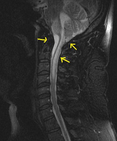
Additional clinical history: Acute leukemia.
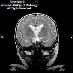
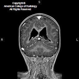
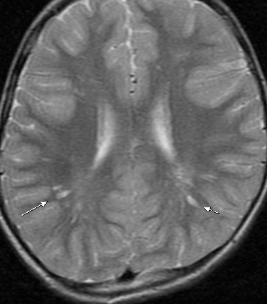

Findings

Large anterior epidural mass extending from posterior clinoid to the cervicothoracic junction measuring approx. 6x3x1.5 cm with mass effect on the anterior pons, medulla, and upper cervical cord. No post-contrast imaging obtained.

Differential diagnosis: Epidural mass
- Metastatic disease
- Lymphoma
- Leukemia/chloroma
- Chordoma
- Osteomyelitis/epidural abscess
- Epidural hematoma
- Primary tumor such as neurofibroma/schwannoma


Diagnosis: Chloroma of epidural space


Key points

AKA granulocytic sarcoma, extramedullary myeloblastoma.
Most commonly occurs in the setting of AML.
Can also occur in setting of chronic myelogenous leukemia and other myeloproliferative disorders.
These tumors can involve any part of the body, either concurrently or sequentially.

Imaging characteristics:
- NECT: Isodense or hyper dense to brain or muscle
- MR: Hypo intense or Iso intense on T1-weighted MR images, heterogeneously Iso intense or hyper intense on T2-weighted MR images
- MR+C: Enhance homogeneously after injection of contrast medium

Paraspinal and intraspinal lesions are also thought to arise from perivenous arachnoid spread of leukemic cells. Uncommonly, spinal involvement by granulocytic sarcoma may cause compression of the spinal cord, cauda equina, or nerve roots

Sunday, March 6, 2011

Service Tax for Diagnostic Centres in India


"In the Union Budget of India for the year 2011-12, the Union Finance Minister spread the service tax net to the Healthcare Sector. All hospitals which are Centrally Airconditioned and have more than 25 beds will have to charge a service tax to the tune of 10%. Similarly all the Diagnostic Centres will also have to charge service tax. "
Is it a fair ask, considering standard healthcare is still out of reach of an average Indian, and it is bound to make things make expensive. Our thoughts are welcome. 

Patient's Prefer- More Speed

In this month American Journal of Roentgenology AJR 2011; 196:605-610 An article by Pat A. Basu et al titled  Creating a Patient-Centered Imaging Service: Determining What Patients Want, according to  author's survey responses,"results needed to be communicated within a few hours for an "acceptable" rating from 95% of patients. Further is concluded by the authors that patients want their results communicated much sooner than is currently practiced. "


Saturday, March 5, 2011

Low-risk of NSF with Dotarem (Gd-DOTA).

In a study presented in ECR 2011  independent prospective study supported by agencies in France have validated the low-risk status of the contrast agent Dotarem (Gd-DOTA).  Detailed report on Diagnostic Imaging

Thursday, March 3, 2011

Miliary tuberculosis-CT

Miliary tuberculosis (also known as "disseminated tuberculosis" is a form of tuberculosis that is characterized by a wide dissemination into the human body and by the tiny size of the lesions (1–5 mm). Its name comes from a distinctive pattern seen on a chest X-ray of many tiny spots distributed throughout the lung fields with the appearance similar to millet seeds—thus the term "miliary" tuberculosis.


Wednesday, March 2, 2011

Post cholecystectomy MRCP


Teaching points

85% of the patients after laparoscopic cholecystectomy become symptom free. However 5% produced symptoms referred to as post cholecystectomy syndrome. The causes could be biliary stricture / Remnant stump calculi / dyskinesia / cystic neuroma / idiopathic.

Laparascopic technique though has been established involves technique which could leave long cystic duct stump. Radiologically when evaluated, this dilated tubular structures in and around the gall bladder fossa is difficult to differentiate whether it is a stump of the duct or remnant of the gall bladder. Long cystic duct stump is defined as more than 1 cm in length. Rogers etal after extensive study, concluded that leaving along cystic duct stump is not risk factor for remnant calculi. Though differences of opinion exists it is now recommended the total excision of the duct is ideal.

Calots triangle ( inferior edge of the liver forming superior border, common hepatic duct medial border and cystic duct inferior border with cystic artery as the contents) contributes to be an important land mark for identification of the cystic duct / gall bladder junction.

This 29 yr old young lady operated 2 years back for cholelithiasis has recurrent abdominal pain especially with fatty foods. This case represent a long tubular dilatation at the end of an apparent convoluted  cystic duct ( measuring approximately 4.5 cm), possibly the remnant gall bladder in an operated cholelithiasis with adhesions. As the patient is periodically symptomatic, re-exploration and excision of the remnant of the gall bladder along with long cystic duct stump is recommended depending  on the progress. Case submitted by Dr MGK Murthy


Tuesday, March 1, 2011

Will Radiologists Become Redundant Ever?

This is a brief conversation i had with a cardiologist friend on facebook who was quite sure that radiologists will become extinct one day and imaging should be done by respective departments themselves. Read on and  post your comments. Reason for my sharing this debate here, spark off a larger debate towards need for sub-specialization in radiology else people will consider us replaceable. Lets learn from this talk see that we can earn respect as specialists when we tell them more than they know already.


Cardiologist Friend : People in every specialty should take over their respective Radiology . Like cardiologists have done. Cardiac Radiology and interventions are our domain. If radiologist does echo cardiogram, we just won't accept , that's it. Same way, why not these Neurologists, Gastro s , Gynaes take over their radiology ? How about that?

Me: i work in speciality neurology-orthopedic centre and i have seen even the best specialists have limitation in interpretation of MRIs so lets keep all diagnostic imaging to us radiologists possibly in future we might just think of sonographers as trained technicians who will do the manual work of scanning as is done world over, leave the highly skilled radiologist for interpretation.

Cardiologist Friend : I am sure neurologists can read neuro images well. Some of my neurology friends have taken up neuro radiology and interventions. I have helped some in carotid interventions too.

Me:  think of it as time spent in learning radiology versus time spent in learning neurology you cannot do both.

Cardiologist Friend : when you learn anatomy, physiology and pathology of neurology better, image is just a part of it. Neurologists read EEG, Nerve conduction etc. They are best suited than some sonographer doing it. For neurologists , it is just a clinical correlation.  Let general surgeons, gastro surgeons and Gastro enterologists do USG. Further we don't allow radiologists do echo or coronary angio at all. Cardiac cath lab is out of bounds for them. We even named cardiac ultra sound differently- Echo cardiography.

Cardiologist Friend :If every speciality takes up their imaging, radiologists will become redundant, in medicine! That's sad! From tomorrow , my radiologist won't offer me tea!

Me: it was a belief years back that radiologists will become redunant , but sadly there will never be a day that radiologists will go out of work,, why dont you suggest that surgenns should see histopath as well?? :).  i work in Neuro hospital and i review all their cases at request and trust me, need is not for neurologists to read cases, but need is for radiologists to sub-specialize and provide quality reads.

Cardiologist Friend : A cooperative work is always better , I think. We co report our CT coronary .

Me : reason is lack of expertise, not vice versa. i know a lot of cardiologists who are at loss when they see cross sections instead of routine coronary images.

Cardiologist Friend :I know some "Giants" who can't distinguish LAD from Lcx in CT.

Me:  ‎:)) this happens as coronary CT is relatively new modality, i know of cardiologists who miss lung metastatis incidentally picked up on cardiac CT

Cardiologist Friend : They need to sit with radiologists to learn, if they are interested.

Me: Same as we sit with cardiologists but that doesnt make us replaceable... :)

Basilar dolichoectasia determining a vascular loop compression syndrome


Findings

Figure 1, Figure 2, Figure 3, Figure 4, and Figure 5: Axial high resolution T2 fiesta images show a dilated and tortuous basilar artery which extends into the left cerebellopontine angle. The visualized inner ear structures are normal.
Figure 6: The basilar artery appears to contact the left trigeminal nerve at the root-exit zone.


Diagnosis: Basilar dolichoectasia


Trigeminal neuralgia is a clinical syndrome composed of paroxysmal facial pain usually confined to the maxillary (V2) and/or mandibular (V3) branches of the trigeminal nerve. Occasionally the opthalmic division (V1) is also affected. This syndrome is more common in patients over the age of 65, with no gender specificity.

VLCS is a recognized cause of trigeminal neuralgia. The offending vessel courses into the anterior cerebellopontine cistern with subsequent irritation of the 5th cranial nerve at the preganglionic root entry zone (REnZ). Additional causes of trigeminal neuralgia include anuersysms, AVMs, and tumors of the cerebello-pontine angle. Demyelinating disorders such as multiple sclerosis are also described as a potential cause.

Thin section high resolution T2 MRI of the CPA/IAC allows the best visualization of the vascular loop. These images also show the anatomic course of the 5th cranial nerve from the root entry zone into meckel’s cave. The imaging protocol should include whole brain T2/FLAIR to exclude additional etiologies such as multiple sclerosis. Axial and coronal T1 of the brainstem with gadolinium enhancement is also helpful to look for cranial neuritis, perineural tumor, and cisternal tumor such as an epidermoid, schwanomma, or meningioma.