Wednesday, November 3, 2004


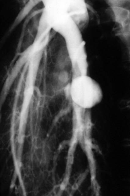
Pulmonary angiography showing a Rasmussen's aneurysm of the right lower lobe incidentally discovered in a patient with a previous history of cavitary tuberculosis of the same lobe.

COPYRIGHT (SUMER)

Tuesday, November 2, 2004


phantom tumour-collection of pleural fluid between lobes, i.e. within the major and minor fissures. The radiographic appearance depends on the shape and orientation of the fissure, the volume of fluid, its position within the fissure and the radiographic projection. Interlobar fluid is particularly common in heart failure.

COPYRIGHT (SUMER)

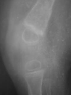
Scurvy
Rare before 6 months of age since the storage of vitamin C in neonate is generally adequate
• Wimberger sign: presence of a sclerotic rim around epiphysis
• White line of frankel:dense zone of provisional calci­fication at the growing metaphysis
• Trumerfeld zone:a lucent zone below white line due to lack of mineralisation
• Pelkan spur:as the area is prone to fractures manifesting at cortical margin
• Osteoporosis
• Subperiosteal haemorrhage.
COPYRIGHT (SUMER)
Tutorials

STRING OF BEADS


PULMONARY EDEMA


BILIARY ATRESIA


MITRAL STENOSIS


MRI IN BLADDER CANCER


RADIOBIOLOGY


COPD X-RAY FINDINGS


WHITAKER TEST


CHYLOTHORAX


PULMONARY INFECTIONS


RADIOSENSTIVITY OF TUMOURS


DYSPHAGIA


ERCP VS MRCP


RADIATION UNITS


SCURVY


Tc99 decay scheme


Monday, November 1, 2004


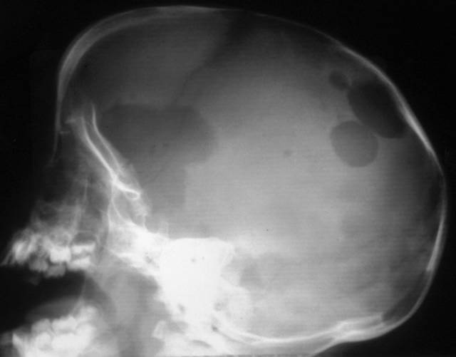
Langerhans' cell histiocytosis

Lateral radiograph of the skull demonstrating multiple lytic lesions of histiocytosis. The anterior lesion has a "geographical" pattern.


COPYRIGHT (SUMER)

imaging news

Portable CT goes to ICU patients


Portable CT scanners can now provide valuable abdominal diagnostic information for intensive care unit patients without forcing them to leave the unit, according to a study in the September issue of the American Journal of Roentgenology.

The first portable CT scanners were deployed at several large institutions in the late 1990s. Little is known, however, about their performance and use in addition to cranial imaging, which remains portable CT's main application. Unlike cranial CT, an abdominal scan requires moving patients from their beds onto the scanning table. But portable scans acquired at the bedside can reduce well-documented risks associated with transport of ICU patients to other hospital locations.

Dr. Michael M. Maher and colleagues at Massachusetts General Hospital retrospectively reviewed spiral portable abdominal CT scans from 107 patients obtained between June 1999 and December 2000. They compared portable CT's image quality and diagnostic value with that of available stationary CT scans.

Although image quality from portable abdominal CT did not match stationary CT, the researchers found they were able to obtain important diagnostic information without moving patients from the ICU.

Researchers obtained 122 portable and 41 stationary CT scans, with 47 and 15, respectively, enhanced by contrast. Intravenous contrast improved portable CT scan quality. Quality scores for portable CT scans, however, were consistently lower than those for stationary CT, both with and without contrast.

Findings on portable CT confirmed 33 conditions suspected before scanning. Portable scanning also detected evidence of infection in 18 patients and hemorrhage in 16, led to seven laparotomies and six percutaneous drainage procedures, and influenced a change in patient management in 33 cases. Surgery or autopsy results confirmed portable CT findings in 12 of 17 cases.

The spiral portable CT scanning protocol included two 285 to 355-mm-long volumes acquired with 5-mm slice thickness during 70 sec at 120 to 130 kVp, 30 to 40 mAs, and a 1 to 1.5-sec pitch. Patient condition and imaging indication determined IV contrast use. Stationary scanning was performed with single- and four-detector spiral scanners.

Interpretation of portable CT studies must proceed with caution, however. Some portable CT parameters, such as accuracy and negative predictive value, were unknown. While portable scanning is useful in many cases, patients who require valuable diagnostic information should be moved to more sophisticated imaging installations whenever possible, researchers said.


Can MRI replace DMSA in the detection of renal parenchymal defects in children with urinary tract infections?

Background: Renal parenchymal defects may be a consequence of urinary tract infections (UTI) in childhood. MRI is a non-radiation imaging modality compared with DMSA scanning. Objective: To compare DMSA with MRI for the detection of renal parenchymal defects in children presenting for radiological investigation after a first UTI. Materials and methods: Both DMSA and MRI were performed at the same appointment in 37 children (aged 4 months-13 years; mean 4.5 years) with a history of UTI. Both planar and SPECT DMSA were performed. MRI of the kidneys employed axial and coronal T1-, T2- and fat-saturated T1-weighted (T1-W) sequences. Some children had imaging after IV contrast medium. Results: The coronal fat-saturated T1-W sequence was the best sequence and it detected all the findings on MRI. MRI had a sensitivity of 77% and a specificity of 87% for the detection of a scarred kidney using DMSA as the gold standard. MRI diagnosed pyelonephritis in two children that had been interpreted as scarring on DMSA. Conclusions: Renal MRI using a single, coronal, fat-saturated T1-W sequence is a rapid, accurate and minimally invasive technique for the the detection of renal scarring that does not employ ionizing radiation.


Pediatr Radiol. 2004 Oct 14 [Epub ahead of print]