"In three cases of cerebral malaria, MR imaging disclosed either cortical infarcts (one case) or hyperintense areas of white matter (two cases) on T2-weighted and fluid-attenuated inversion-recovery sequences. These white matter abnormalities were, in one case, sharply limited, symmetrical, hyperintense, and unenhanced; in the other case, they were diffuse, hyperintense, and had a more limited focus. The diffuse hyperintensity was probably due to edema, whereas focal lesions were probably associated with gliosis."
Saturday, July 15, 2006
Cerebral Malaria-MRI
MR of cerebral malaria. In AJNR Am J Neuroradiol 1998 May;19(5):871-4. By Cordoliani YS et al.

Renal Artery Stenosis-Anatomical Distribution
Vo NJ et al in their article entitled "Anatomic distribution of renal artery stenosis in children: implications for imaging" in Pediatr Radiol 2006 Jul 4; [Epub ahead of print] found that-
"Focal Renal Artery stenosis in their study group was distributed as follows 25% main renal artery, 50% 2nd order branch, 12.5% 3rd order branch, and 12.5% accessory renal artery. They concluded Hypertensive children without comorbid conditions who have RAS usually have single, focal branch artery stenoses. This distribution supports angiography in these patients because of its superior sensitivity in detecting branch vessel disease and its therapeutic role in percutaneous transluminal renal angioplasty."
Tuesday, July 11, 2006
Some useful Radiology RSS Feeds
Found some useful Radiology Related RSS feed which Radiologists can add to their RSS readers and keep updated to the latest in Radiology.
Sumer's Radiology Site
http://sumerdoc.blogspot.com/atom.xml
Journal of Computer Assisted Tomography
http://www.jcat.org/pt/re/jcat/toccurrentrss.xml
Topics in Magnetic Resonance Imaging
http://www.topicsinmri.com/pt/re/tmri/toccurrentrss.xml
Journal of Thoracic Imaging
http://www.thoracicimaging.com/pt/re/jti/toccurrentrss.xml
Medscape Radiology Headlines
http://www.medscape.com/cx/rssfeeds/radiology.xml
Sumer's Radiology Site
http://sumerdoc.blogspot.com/atom.xml
Journal of Computer Assisted Tomography
http://www.jcat.org/pt/re/jcat/toccurrentrss.xml
Topics in Magnetic Resonance Imaging
http://www.topicsinmri.com/pt/re/tmri/toccurrentrss.xml
Journal of Thoracic Imaging
http://www.thoracicimaging.com/pt/re/jti/toccurrentrss.xml
Medscape Radiology Headlines
http://www.medscape.com/cx/rssfeeds/radiology.xml
Friday, July 7, 2006
Availability of Sonography in Emergency Gynecology Unit useful
In an Article entitiled "Impact of the availability of sonography in the acute gynecology unit." in Ultrasound Obstet Gynecol 2006 Jun 29; [Epub ahead of print] by Haider Z et al, the authors investigated the impact of the availability of transvaginal sonography at the time of initial assessment of the emergency gynecology patient. The initial assessment of acute gynecology patients is usually based on history and clinical examination and does not involve ultrasound. Authors found that Following the ultrasound examination there was a change in clinical management for 38.1% of non-pregnant women and a reduction in admissions (from 37.1% to 19.4%) and outpatient follow-up examinations (from 25.7% to 18.1%). It appears that the availability of transvaginal sonography at the time of initial assessment of emergency gynecology patients improves diagnostic accuracy and reduces unnecessary admissions and follow-up examinations.
Interesting post on Desert Imaging Blog
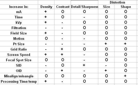 + = increase, - = decrease, O = no change
Interesting post in Desert Imaging in which he talks about a Real Tough Radiography examination he appeared.
He goes like-
"It was one of those tests with questions like:"If you increase focal spot size how does it affect image detail?", "If kVP is increased by 15% what will happen to image density?", "When you increase OID what happens to radiographic contrast?" yada, yada, yada, blah, blah, blah, etc., etc., etc......."
And then follows up with a very useful Table for all Radiographers as well as Radiology Residents...
Thursday, July 6, 2006
Overdiagnosis Of Breast Cancer
Overdiagnosis and Overtreatment of Breast Cancer: Is Overdiagnosis an Issue for Radiologists?
Reference-Breast Cancer Res. 2006;8(2)
Complete article here at Medscape
"Overdiagnosis is diagnosis of cancers that would not present within the life of the patient and is one of the downsides of screening. This applies to low-grade ductal carcinoma in situ and some small grade 1 invasive cancers. Radiologists are responsible for cancer diagnosis, but at the time of diagnosis they cannot determine whether a particular low-grade diagnosis is one to which the definition of overdiagnosis applies. Overdiagnosis is likely to be driven by technological developments, including digital mammography, computer-aided detection and improved biopsy techniques. It is also driven by the patient's fear that cancer will be missed and the doctor's fear of litigation. It is therefore an issue of importance for radiologists, presenting them with difficult fine-tuned decisions in every assessment clinic that are ultimately counted later by those who evaluate their screening."
Monday, July 3, 2006
Future of Double Contrast Barium Enema in Question
In a Study by Ferrucci JT, Double-contrast barium enema: use in practice and implications for CT colonography in AJR Am J Roentgenol 2006 Jul;187(1):170-3
"This study examines the use and yield of double-contrast barium enemas (DCBEs) for colorectal polyp detection in current clinical practice outside the research setting. A total of 244 out of 665 (36.7%) barium enema studies were performed using the double-contrast technique over the 4-year period-that is, approximately one per week. The most common indication for a DCBE (109/244 or 44.6%) was to complete a failed, incomplete, or inconclusive colonoscopy.Overall, only 14 of the 244 (5.7%) studies gave positive reports for polyps, and of these, five were shown to be false-positive at later colonoscopy. Only six polyps 10 mm or larger were positively detected during the entire study, which is approximately one per 60 studies or one every 8 months
Authors concluded that in their centre a DCBE is a low-yield procedure for detecting polyps, with a high false-positive rate, and is not likely to be performed by experienced practitioners in the future."
Subscribe to:
Posts (Atom)