Sunday, October 31, 2004


Sequestrum- A large rod-like sequestrum is observed in the medullary canal owing to chronic osteomyelitis.

COPYRIGHT (SUMER)

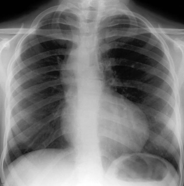
Chest radiograph demonstrates decreased pulmonary vascularity, normal cardiac size, concave main pulmonary artery segment and right aortic arch... the boot shaped heart..or cor-en-sabot..

COPYRIGHT (SUMER)

Saturday, October 30, 2004

STRING OF BEADS

string of beads in radiology is a sign which has been described for various pathologies.. it is seen in--


String of beads in radiology-

  • Fibromuscular dysplasia

  • Chr. Pancreatitis (chain of lakes)
  • Small bowel obstruction
  • varicose bronchiectasis


Fibromuscular dysplasia

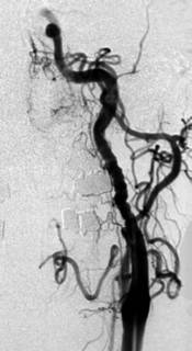
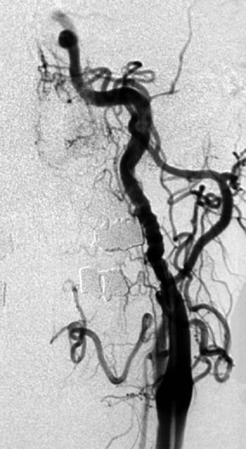
condition of unknown aetiology that involves the extracranial internal carotid arteries and vertebral arteries. The internal carotid is involved in about 75% of cases and the vertebral in less than 25%. It is characterized by narrowing of the affected vessel with a string of beads appearance , due to focal annular repetitive intimal and medial proliferative changes. Not infrequently an incidental finding, fibromuscular hyperplasia may be a cause of dissection and is associated with an increased incidence of intracranial aneurysms.
COPYRIGHT (SUMER)

Pulmonary edema

Pulmonary oedema


The radiographic changes of hydrostatic oedema are quite characteristic. In the normal adult, the lower lobe pulmonary vessels are larger than the upper lobe vessels due to gravitational forces. As the left-sided pressure increases, the blood is diverted to the upper lobes. This results in "cephalization" with the upper lobe vessels becoming larger than the lower lobe vessels. As left heart pressure increases, fluid enters the peribronchovascular interstitium. As the interstitium becomes oedematous, the interlobular septa become prominent and the markings indistinct.  Pleural effusions are frequent in the more severe stages of left heart failure with a slight predominance to the right.

Thursday, October 28, 2004


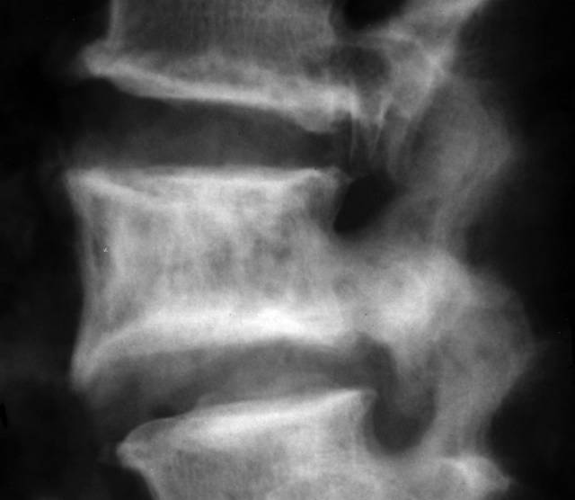
Lateral radiograph of a lumbar vertebral body demonstrates the condensation of the bone along the periphery of the expanded vertebral body which has been termed a "picture frame" appearance
classical of pagets disease
COPYRIGHT (SUMER)

Paget's disease
Lateral radiograph of the skull demonstrates a thickened calvarium and fluffy radiodense regions which is referred to as a "cotton-wool" appearance.

COPYRIGHT (SUMER)