Saturday, May 28, 2005

Journal Club-The value of high-field MRI (3T) in the assessment of sellar lesions.

Pinker K, Ba-Ssalamah A, Wolfsberger S, Mlynarik V, Knosp E, Trattnig S.
The aim of this study was the evaluation of the normal sellar anatomy in vitro and in vivo with high-field MRI and its application in the diagnosis of sellar pathologies in comparison to standard MRI. All high-field MR images were obtained using a 3T Bruker Medspec 30/80 Scanner with a head birdcage transmit/receive coil and an actively shielded gradient system with a maximum gradient strength of 45mT/m. Firstly an in vitro study of the sella turcica was performed to depict normal pituitary and sellar anatomy at high field. After a pilot-study this sequence-protocol was established: A RARE sequence (TR/TE = 7790/19ms; matrix size, 512 x 512; RARE factor = 8, FOV, 200mm) was used for T2-weighted coronal, axial and sagittal images. A 3D gradient echo sequence with magnetization-preparation (MP-RAGE, TR/TE/TI = 33.5/7.6/800ms, matrix size, 512 x 512; FOV, 200mm, effective slice thickness, 1.88mm; 3 averages) was used for acquisition of T1-weighted pre- and post-contrast images. Between January 2002 and March 200458 patients were enrolled in this study. Seven patients were examined for suspected microadenoma and in 51 patients 3T MRI was used to obtain additional information about the sellar lesion already known to be present from standard MRI. In 21 cases the accuracy of the imaging findings was assessed afterwards by comparison with intraoperative findings. The infiltration of the medial cavernous sinus wall was suspected on standard MRI on 15 sides (47%), on high-field MRI on 9 sides (28%) and could be verified by intraoperative findings on 6 sides (19%). Accordingly, sensitivity to infiltration was 83% for 3T and 67% for standard MRI. Specificity was 84% for 3T and 58% for standard MRI. Moreover, high-field MRI revealed microadenomas in 7 patients with a median diameter of 4mm (range 2-9mm). The segments of the cranial nerves were seen as mean 4 hypointense spots (range 2-5 spots) on high-field MRI in contrast to 3 spots (range 0-4 spots) on standard MRI. This difference was considerably significant (P <>High-field MRI is superior to standard MRI for the prediction of invasion of adjacent structures in patients with pituitary adenomas and improves surgical planning of sellar lesion.
Eur J Radiol. 2005 Jun;54(3):327-34.


Saturday, May 21, 2005

Journal watch-Bone measurements of infants in the first 3 months of life by quantitative ultrasound:influence of gestational age, season&postnatal age

Liao XP, Zhang WL, He J, Sun JH, Huang P.
Background: There are a few quantitative ultrasound (QUS) studies of bone status for Chinese children.
Objective: To evaluate the clinical application and to investigate the bone status of neonates and young infants with QUS.
Materials and methods: An ultrasound bone sonometer was used to measure the bone speed of sound (SOS) of the tibia in 542 neonates within 3 months of birth.
Results: At birth, no significant difference of SOS was found between boys and girls, but there was a significant difference of SOS between premature infants and full-term infants. The SOS in neonates born during spring and summer was significantly lower than those born during autumn and winter. There were significant correlations between SOS and gestational age, and between bone SOS and birth weight in appropriate for gestational age (AGA) infants. Multiple regression analysis found that gestational age and infant birth season were two important factors influencing SOS. During the first 3 months, there was no significant difference in SOS between sexes. The SOS of infants showed an inverse correlation with postnatal age, and the decrease of bone SOS with age in premature infants was more marked than in full-term infants.
Conclusions: QUS is suitable for evaluating bone status in infants with high precision. The study offers some basic data for neonates and young infants
Pediatr Radiol. 2005 May 12; [Epub ahead of print]

Tuesday, May 17, 2005

AIIMS MAY 2005 FULLY SOLVED FINALLY AVAILABLE


Author-
Dr Sumer Kumar Sethi
MBBS (MAMC), MD Radiodiagnosis (LHMC) (Gold medalist)
CURRENTLY SENIOR RESIDENT RADIODIAGNOSIS LHMC
ONE OF THE TOPPERS IN AIPG 2000, AIIMS 2000 AND PGI 2000. EARLIER WAS IN TOP 5 IN CBSE PMT AND DPMT. AUTHOR OF "REVIEW OF RADIOLOGY" & MAY 2004 AIIMS, AIIMS NOV 2004, ALL INDIA 2005 BOOK-POPULAR TEXTS FOR PG ASPIRANTS.

Dr Sidharth Sethi

MBBS (MAMC), MD Pediatrics (STD)


Salient features of the book
· SUBJECTWISE EXPLANATION OF THE AIIMS MAY 2005 PAPER
· EASY EXPLANATIONS WITH SUITABLE DIAGRAMS
·EVEN THOUGH IT IS COMPREHENSIVE ITS STILL MANAGEABLE IN SHORT TIME AVAILABLE TO STUDENTS.
· USEFUL FOR ALL INDIA, AIIMS, PGI AND VARIOUS STATE PG EXAMS
· ALL INFORMATION CHECKED IN STANDARD AND EASILY AVAILABLE TEXT BOOKS
· AN IMPORTANT PART OF PREPARATION FOR PG ENTRANCE GIVEN THE HIGH NUMBER OF REPEATS IN VARIOUS EXAMINATIONS
·FIRST BOOK TO OFFER ONLINE SUPPORT AT www.sumerdoc.blogspot.com AND www.indianradiology.com
COPYRIGHT (SUMER)

Monday, May 16, 2005

Journal watch-Comparison of recall and cancer detection rates for immediate versus batch interpretation of screening mammograms.

Ghate SV, Soo MS, Baker JA, Walsh R, Gimenez EI, Rosen EL.
PURPOSE: To retrospectively compare recall and cancer detection rates between immediate and subsequent batch methods for interpretation of screening mammograms.
MATERIALS AND METHODS: Institutional review board approval was obtained, and informed consent was waived. Retrospective analysis was performed for 8698 screening mammograms obtained between January 1 and October 31, 2001, which were interpreted either immediately (n = 4113) or subsequently with batch method (n = 4585). Data were collected from data reporting system and patient billing records. Patients with high risk factors were excluded; 3441 patients were in the immediate group, and 3932 were in the batch group. The two groups were compared with respect to age, breast density, and availability of comparison films with Wilcoxon rank sum test. Recall rates and cancer detection rates for each group were determined and compared with Pearson chi(2) test; false-negative rates were compared with Fischer exact test.
RESULTS: A significant difference (P < .001) was noted in recall rates between immediate (18%) and batch (14%) groups; however, no significant difference (P = .7) was noted in cancer detection rates (immediate, 0.5%; batch, 0.4%). Mean age of patients was 56.8 years (age range, 21-96 years) in the immediate group and 56.2 years (age range 24-98 years) in the batch group (P = .02). Comparison of breast densities between groups indicates no statistically significant difference (P = .4). The batch group had significantly fewer comparison mammograms (3106 [79%]) available than the immediate group (2856 [83%]) (P < .001). There was no significant difference in false-negative rates between the immediate group (0.1%) and the batch group (0.1%) (P > .99).
CONCLUSION: Immediate interpretation of screening mammograms resulted in a statistically significant increase in recalls and additional clinical work-ups of perceived abnormalities; however, no significant difference in cancer detection rate was detected between groups.
Radiology. 2005 Apr;235(1):31-5.

Thursday, May 12, 2005

IMAGE CASE-Tuberculosis of the female genitourinary tract


A gynecological US showing evidence of endometrial calcification and left hydrosalpinx-Tubercular etiology
COPYRIGHT (SUMER)

Saturday, May 7, 2005

A follow up to the Image case-About testicular microlithiasis

Testicular microlithiasis: a review and its association with testicular cancer.
Rashid HH, Cos LR, Weinberg E, Messing EM
Testicular microlithiasis (TM) is an entity of unknown etiology that results in the formation of intratubular calcifications. It is of concern to the urologist because of its possible association with intratubular germ cell neoplasia and testicular germ cell cancer. Although commonly present in patients with germ cell tumors, there appears to be no definitive association with TM and cancer. Therefore, follow-up at this time should be dictated based on risk factors for developing testis cancer more than on the presence of TM.
Urol Oncol. 2004 Jul-Aug;22(4):285-9.

Thursday, May 5, 2005

Image Case


An Interesting Case


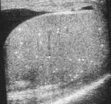
A Gray Scale Ultrasound image of testes showing multiple hyperechoic foci-
A case of Testicular Microlithiasis

COPYRIGHT (SUMER)