Select posts by Sumer Sethi, the author of the online Radiology magazine Sumer's Radiology Site will now also be available in The Medical Blog Network
Link- http://www.healthvoices.com/blog/sumersethi
Wednesday, January 11, 2006

Friday, January 6, 2006
Random walks in old manila
One of my hobbies in life is to walk on late afternoons in old Manila. I particularly love this time of the day, appreciating how the light of day begins to darken to welcome the lovely hue of the comign evening. No, no. This is not an old photo. I just shot this lovely Intramuros gate in sepia in order to get a feel of what it was like during the olden days.
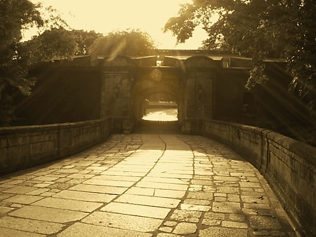 No, no. This is not an old photo. I just shot this lovely Intramuros gate in sepia in order to get a feel of what it was like during the olden days.
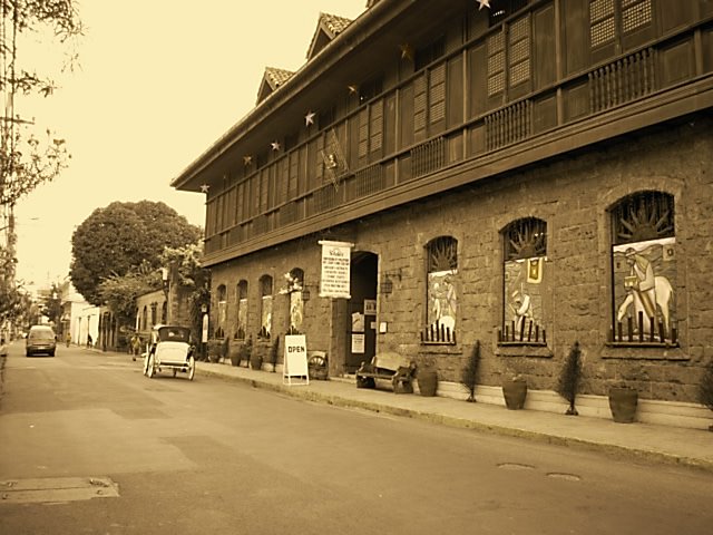 This is El Amanecer, along General Luna ST., in Intramuros. This imposing colonial edificio houses an art gallery, an antique shop, and a filipiniana bookshop. A haven for a vintage lover like me. Then I headed over to Manila Bay, because I wanted to watch the world famous Manila sunset.
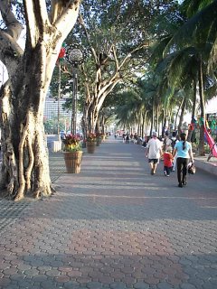
The baywalk, a fresh breeze welcomed me as I walked on its tiled walkway. 4:30pm
To catch the lovely hues in the sky as the sun drips under the horizon.
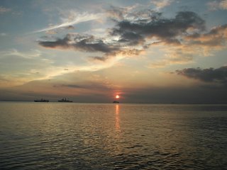
When it was quite dark, I headed over to the ancient Malate Church to catch its radiant beauty against a darkening sky.
This is life. I enjoy it while I'm still young. When I get old I may not be strong enough to walk and enjoy my Manila.
There are several bars along Nakpil and Mabini streets. I entered in one for my dinner and a few relaxing drinks.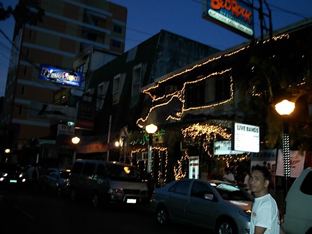
Tuesday, January 3, 2006
Vote for my site, Polls for 2005 Medical Weblog awards are open!!
Dear Friends and Readers
As you all know your favourite Sumer's Radiology Site has been nominated in three categories in 2005 Medical Weblog Awards..
So please support this site by voting in all three links given below...
So please support this site by voting in all three links given below...
PLEASE NOTE VOTING IS REQUIRED IN EACH CATEGORY SEPERATELY
Monday, January 2, 2006
Calcaneal Fractures-Role of CT
Fractures of the Calcaneus: A Review with Emphasis on CT
RadioGraphics 2005;25:1215-1226
Aditya Daftary, MB, BS, Andrew H. Haims, MD and Michael R. Baumgaertner, MD
"The calcaneus is the most commonly fractured tarsal bone and accounts for about 2% of all fractures. Advances in cross-sectional imaging, particularly in computed tomography (CT), have given this modality an important role in identifying and characterizing calcaneal fractures. Fracture characterization is essential to guide the management of these injuries. Calcaneal fractures have characteristic appearances based on the mechanism of injury and are divided into two major groups, intraarticular and extraarticular. Most calcaneal fractures (70%–75%) are intraarticular and result from axial loading that produces shear and compression fracture lines. Of the two major systems for classifying intraarticular fractures–Hannover and Sanders–the latter is used most often and is helpful in treatment planning and determining prognosis. Extraarticular fractures account for about 25%–30% of calcaneal fractures and include all fractures that do not involve the posterior facet. Familiarity with calcaneal anatomy and fracture patterns is essential for radiologists to guide the treating physicians. "
Read the full article here-
Friday, December 30, 2005
Sumer's Radiology Site nominated for following categories in Annual Medical Weblog awards

The second annual Medical Weblog Awards! These awards are designed to honor the very best in the medical blogosphere, as decided by the readers of these fine medical blogs.
It's been another year filled with explosive growth, stirring debate, and excellent writing -- in a number of fields. Sumer's Radiology Site has been nominated in following categories. Sumer's Radiology Site is the only weblog to be nominated in three categories!! Thanks to all readers and visitors...
-- Best Medical Weblog
-- Best Clinical Sciences Weblog
-- Best Medical Technologies/Informatics Weblog
Click here to see the other nominees
Please note-Polls will be open from Tuesday, January 3, 2006 and will close at midnight on Sunday, January 15, 2005 (PST).
I ll put up the link shortly...
Image Quiz-GIT

Image Quiz
A 38 year old woman presented with dyspepsia. A Barium swallow was done. What is the diagnosis?
Answer-Sliding Hiatus Hernia
Winners-Jon Mikel, Sharon, Alireza, K Sudhakar
Leave your answers in the comments section. The correct answer and winners will be published on the site next week.
COPYRIGHT (SUMER)
Monday, December 26, 2005
Ultrasound in Ascariasis
Ascaris lumbricoides is a common nematode infesting a major percentage of human beings worldwide. It grows to a maximum length of 35 cm. This species is host specific to human beings and lives longer (1-2 years) with in the small intestine.
However, as the bowel loops are just under the abdominal wall, the detection of intestinal ascariasis is difficult by routine abdominal probes. It demands the use of a higher frequency high-density probe of 5 - 10 MHz. The live worm on longitudinal section appears as a writhing tubular shadow having brighter margins described by some as 'strip sign'. There is a hypoechoic core producing the 'inner tube sign'. The coiled worm appears as 'spaghetti'. The cross-sectional picture is also characteristic of a tubular body described as the ring sign or bull's eye sign if seen with in the CBD or a narrow lumen.
Full article with images here(Full free text available)-
K BALAKUMAR
Ind J Radiol Imag 2005 15:1:107-108
Subscribe to:
Posts (Atom)