Wednesday, January 17, 2007

IUCD and MRI

"When patients with an implanted contraceptive device undergo MRI, it must be ensured that the examination involves no risk to the patient (MR safety) and that the diagnosis is not affected by artifacts or the function of the device compromised (MR compatibility). Two basic types of intrauterine devices can be distinguished: the metal-containing/metal-free intrauterine device (IUD) and the hormone-containing implant, the fully metal-free intrauterine system (IUS), as well as the ESSURE insert made of stainless steel, which has been approved for use in Europe since February 2001.The metal-containing and metal-free IUDs and ESSURE are MRI compatible up to a magnetic field strength of 1.5 T. They do not interact in any relevant way with the external magnetic or high-frequency field and the temperature increase is within the physiologic range. The implants merely produce a local signal void with a shape that depends on their orientation relative to the magnetic field lines. At 3 T, only the metal-free IUD and the IUS are MRI safe in terms of the material used. In contrast, metal-containing IUDs and the ESSURE have not yet been fully evaluated in the 3 T field, which is why they represent a contraindication to MRI. No data are available on the MRI compatibility at 3 T for any of these devices"
Reference-
How safe is magnetic resonance imaging in patients with contraceptive implants?
Radiologe 2006 Jul;46(7):574-8.

Are CXR in preemployment check up justified?

The legal and medical basis for chest radiographs as part of pre-employment examinations (PEE) at a University Hospital is evaluated. The radiographs are primarily performed to exclude infectious lung disease. A total of 1760 consecutive chest radiographs performed as a routine part of PEEs were reviewed retrospectively. Pathologic findings were categorized as "nonrelevant" or "relevant." No positive finding with respect to tuberculosis or any other infectious disease was found; 94.8% of the chest radiographs were completely normal. Only five findings were regarded as "relevant" for the individual. No employment-relevant diagnosis occurred. The performance of chest radiography as part of a PEE is most often not justified. The practice is expensive, can violate national and European law, and lacks medical justification.
Are chest radiographs justified in pre-employment examinations? Presentation of legal position and medical evidence based on 1760 cases.
Radiologe 2006 Jul;46(7):567-73

CT Angiography and Cancer Risk

"As the number of cardiovascular CT angiography scans continues to grow, researchers are attempting to gauge the potential effects of radiation exposure during the procedure. Although extensive epidemiologic studies are not yet available, new research suggests a measurable risk. A study presented at the American Heart Association meeting by Andrew J. Einstein of the Mount Sinai School of Medicine in New York City found that a single cardiovascular CTA study produced a non-negligible lifetime attributable risk (LAR) of cancer in patients. The risk was greatly influenced by factors such as the patient's age and gender, as well at the scan protocols used."

Reference and full article on Diagnostic Imaging, Study finds measurable cancer risk with CTA

Warthin's tumor


Findings

Figure 1: Contrast enhanced axial CT scan image showing the predominantly cystic left intraparotid mass, with enhancing eccentric nodule and minimal wall thickening. Right parotid gland is normal. No cervical lymphadenopathy was seen on the study.

Differential diagnosis:
- Pleomorphic adenoma
- Warthin’s tumor
- Lymphoma
- Lymphoepithelial cyst
- Mucoepidermoid carcinoma
- Adenoid cystic carcinoma
- Metastasis


Diagnosis: Warthin's tumor


Also known as papillary cystadenoma lymphomatosum, Warthin’s tumors exhibit a slight male predominence and most commonly occur in patients older than 50. The majority of cases are seen in patients in their seventh to eighth decade of life. They are the second most common benign parotid tumor next to pleomorphic adenoma (approximately 75% to 80% of benign parotid tumors are pleomorphic adenoma). Smokers have an eightfold increased risk of developing this tumor, which likely accounts for the historic male predominance. Warthin's tumors are the most common bilateral parotid tumors. About 6% to 10% of patients will have bilateral tumors. They most commonly arise in the tail of the parotid gland, usually presenting as painless and slow-growing masses. There is a very low incidence of malignant transformation.

Typical CT characteristics include a small, ovoid, well-circumscribed, smoothly marginated mass of the posterior parotid gland. The tumor can be cystic or lymphoma-like in appearance. Warthin’s tumors often present as homogeneous soft tissue densities. Cyst formation is common, and the classic appearance is of a cystic lesion with focal tumor nodule(s). On T1-weighted MR, the mass contrasts to the hyperintense signal of the normal parotid gland. On T2WI, Warthin’s tumors are often heterogeneous, with variable signal intensity. Warthin’s tumors show significant uptake of Technetium 99m pertechnetate, as does oncocytoma.

Warthin’s tumor is usually treated by surgical excision. Facial nerve involvement is rare and the facial nerve can usually be spared. Conservative management with close follow-up is acceptable, particularly in poor surgical candidates.

Saturday, January 13, 2007

Polyorchidism-A Case Report


Polyorchidism:
The presence of more than 2 testes is called polyorchidism. It is rare and less than 100 cases are reported in medical literature.Dr Joe presents a case of polyorchidism. The patient presented with a small, painless mass in the left scrotum. Physical examination revealed a small, non-tender lump below the left testes. Sonography revealed a well-defined, solid ovoid mass of less than 2 cms., just below the left testes. It shows same echogencity and echotexture as the testes. This is a very important point that clearly identifies the mass as a supernumerary testes.
D/d: this includes bilobate testes; other differentials include solid masses like fibroma, lipoma and neurofibroma.
Prognosis: polyorchidism is associated with higher risk of malignancy and torsion of the supernumerary testes. Normal spermatogenesis has been reported in the extra testes. Careful follow up examination of the testes is usually sufficient.

Case submitted by-
DR. JOE ANTONY, MD,
Consultant Radiologist.

free to view ultrasound gallery
http://drjoea.googlepages.com


Healthcare in Cochin and more
http://cochinblogs.blogspot.com

Thursday, January 11, 2007

Trigeminal schwannoma


Findings

CT postcontrast reveals an unenhancing soft tissue mass centered at Meckel's cave on the left (Figure 1)
CT bone window (Figure 2) reveals a markedly enlarged foramen ovale on the left, with smooth, corticated margins.
Axial T1-WI (Figure 3) demonstrates a well-circumscribed extraaxial mass in the region of left Meckel’s cave that is mildly hypointense to brain parenchyma. The mass shows heterogeneous signal intensity on FLAIR (Figure 3). Postcontrast images show moderately intense enhancement, extending into the cisternal segment of cranial nerve V (Figure 4). The mass is seen to extend through a widened foramen ovale into left infratemporal fossa and present as masticator mass (Figure 4).


Diagnosis: Trigeminal schwannoma


Schwannomas account for 6% to 8% of intracranial neoplasms. Vestibular schwannomas are the most common cranial nerve schwannomas, followed by trigeminal and facial schwannomas.

The trigeminal nerve (CN V) exits the brainstem at the level of the mid pons, and its three divisions—the ophthalmic (CN V1), maxillary (CN V2), and mandibular (CN V3) branches—together proceed anteriorly toward the trigeminal ganglion in the Meckel’s cave. From here, the mandibular division exits inferiorly via the foramen ovale. The maxillary and ophthalmic divisions continue anteriorly along the lateral aspect of the cavernous sinus. Eventually, the ophthalmic division enters the orbit via the superior orbital fissure, while the maxillary division exits the cranial vault through the foramen rotundum.

Trigeminal schwannomas are encapsulated tumors of Schwann cells displace rather than infiltrate fascicles of CN V3.

Trigeminal schwannomas usually present in the third to fourth decade with sensory disturbances. Mastication muscles weakness or facial pain is less common.

Trigeminal schwannomas may arise along any segment of CN V, but the majority develop at the level of trigeminal ganglion. They may then extend posteriorly into the posterior fossa, or anteriorly through the skull base foramina, and often have a “dumbbell” configuration.

On nonenhanced CT scans, most schwannomas are isodense relative to brain parenchyma. Calcification or areas of hemorrhage are rare. On contrast-enhanced CT scans, the enhancement pattern is typically homogeneous. Foramen ovale is most commonly enlarged by CN V3 schwannoma.

Schwannomas typically are isointense or slightly hypointense relative to gray matter on T1-weighted images and hyperintense on T2-weighted images. Gadolinium enhancement typically is homogeneous, although larger schwannomas can show areas of cystic degeneration and heterogeneous signal intensity. Atrophy of masticator muscles may be seen as high signal fatty infiltration on T1-weighted images with volume loss.

Surgical resection is the treatment of choice.

Trigeminal schwannoma may be associated with Neurofibromatosis type 2, especially if it occurs in younger patients.

Tuesday, January 9, 2007

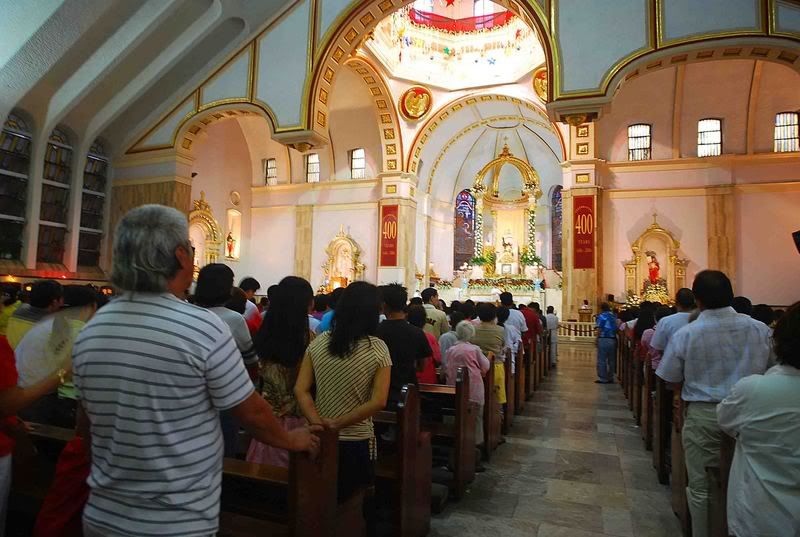
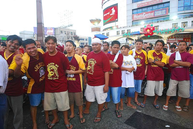
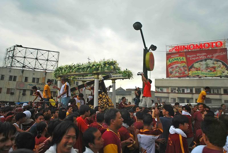
Ika-400 Taon na Kapistahan ng Kiyapo

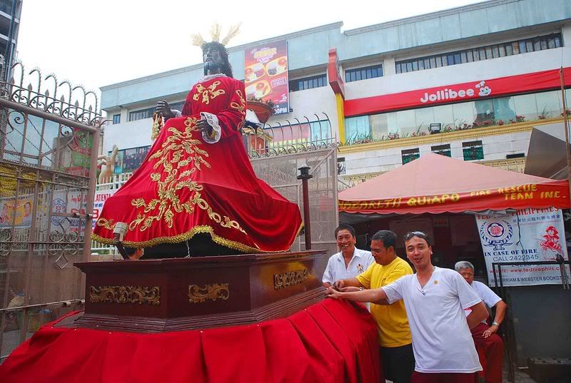
The Black Nazarene being prepared for the procession


Quinta Market near the Quiapo Church


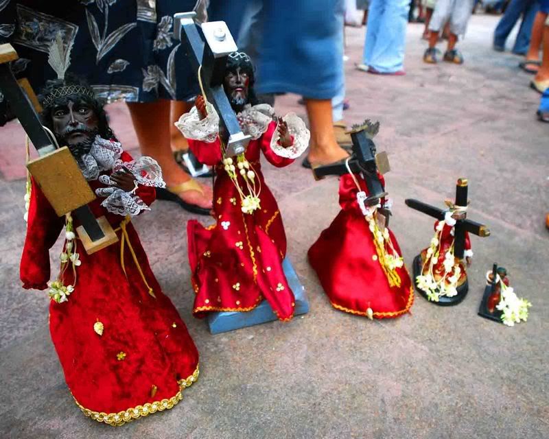
A devotee's collection of Nazareno statuettes in various sizes


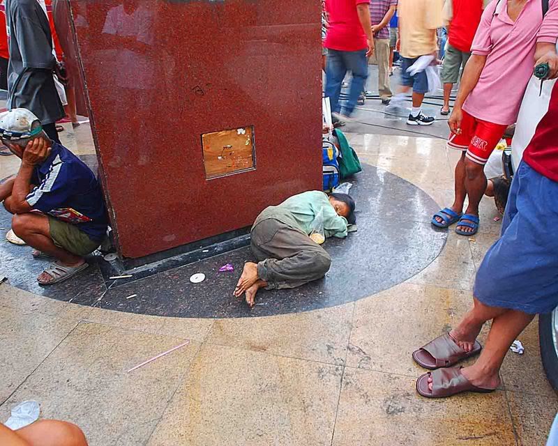
A sleeping "pulubi" in front of the Quiapo Church. This pulubi is unmindful of the massive crowd surrounding her.


A vendor of Nazarene shirts


George Tapan and me, (with two friends) chat while waiting for the procession to start


The crowd gathers in front of Quiapo Church hoping to catch a glimpse of the Nazareno


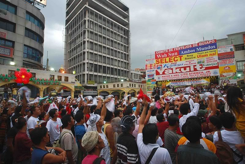
Devotees wave their towels to greet the Black Nazarene amidst shouts of "Viva!"


Plaza Miranda, Quiapo Manila


The mass prior to the procession


Devotees form themselves into a brigade to surround the Nazarene and touch their white towels to him.


The massive crowd surround the Black nazarene to get their towels touch the holy image.