Friday, December 31, 2004


HAPPY NEW YEAR 2005 TO ALL!!!
COPYRIGHT (SUMER)

Gods of Earth and Sea.

And the old lady cries.

And the lonely children suffer.

Their mother's now alone.

Their fathers wont come home.


And the old lady cries.

And the old lady cries.


The gods were hungry.

And the gods weren't happy.

They had offered their jewellery

They had paid and prayed only.


But the stones know no mercy.

Their hunger must be appeased.

While the guilty run free.

The innocent are punished.


But the little girl asks.

Why were we punished?

What did we do wrong?

We would have followed

Had we been told


And the old lady cries.

Her son will not return.

Her faith will not rise

The stones, she will burn.


And the anger wont subside.

Like the waves killed her love.

Her revenge will come.

Her children she must save.

But her anger wont subside
.


Darker-Side-Of-Saturn

Thursday, December 30, 2004


USG image showing ascariasis or a round worm in a bowel loop!!!
COPYRIGHT (SUMER)

Monday, December 27, 2004

Disaster strikes South-east Asia

USGS: Warnings Could Have Saved Thousands in Asia

A warning center such as those used around the Pacific could have saved most of the thousands of people who died in Asia's earthquake and tsunamis, a U.S. Geological Survey official said on Sunday.

None of the countries most severely affected -- including India, Thailand, Indonesia and Sri Lanka -- had a tsunami warning mechanism or tidal gauges to alert people to the wall of water that followed a massive earthquake, said Waverly Person of the USGS National Earthquake Information Center.

"Most of those people could have been saved if they had had a tsunami warning system in place or tide gauges," he said.

"And I think this will be a lesson to them," he said, referring to the governments of the devastated countries.

Person also said that because large tsunamis, or seismic sea waves, are extremely rare in the Indian Ocean, people were never taught to flee inland after they felt the tremors of an earthquake.

Tsunami warning systems and tide gauges exist around the Pacific Ocean, for the Pacific Rim as well as South America. The United States has such warning centers in Hawaii and Alaska operated by the U.S. Geological Survey. But none of these monitors the Indian Ocean region.

The 8.9-magnitude underwater quake -- one of the most powerful in history -- off the Indonesian island of Sumatra devastated southern Asia and triggered waves of up to 30 feet high.

U.S. seismologists said it was unlikely the Indian Ocean region would be hit any time soon by a similarly devastating tsunami because it takes an enormously strong earthquake to generate one.

"That's really what has created all of these problems -- is that the earthquake is just so massive," said Dan Blakeman, a USGS earthquake analyst.

But Person said governments should instruct people living along the coast to move after a quake. Since a tsunami is generated at the source of an underwater earthquake, there is usually time -- from 20 minutes to two hours -- to get people away as it builds in the ocean

FROM-WASHINGTON (Reuters)


Antenatal USG at 18 weeks showing bilateral enlarged echogenic kidney with oligohydramnios!! suggestive of autosomal recessive polycystic kidneys!!!
COPYRIGHT (SUMER)

Friday, December 24, 2004

Thursday, December 23, 2004


Ultrasound abdomen transverse section showing cystic dilatation of distal common bile duct suggestive of choledochal cyst!!!
COPYRIGHT (SUMER)

Monday, December 20, 2004

Medical news

Report from RSNA: CTA nails down role of gold standard for pulmonary embolism

Whether it scans in four slices or 16, CT is the method of choice for detecting pulmonary emboli, researchers said at the RSNA meeting. Even four-slice technology boasts a negative predictive value greater than 99%, better than ventilation/perfusion (V/Q) studies or conventional pulmonary angiography.

Differences among four-, eight-, and 16-slice scanners appear marginal so far, but few data are available to solidify any claims of superiority for 16, except in speed. Researchers at Stanford University clocked lung scan times at 26.6 seconds for four-slice, 9.25 seconds for eight-slice, and 5.45 seconds for 16.

Dr. Alessandro Napoli and colleagues at Stanford reviewed CT studies for 1240 consecutive patients referred for suspected pulmonary emboli. Overall, 20% of cases were deemed positive, with 41% of emboli found in the segmental arteries, 27% in lobal regions, 17% in subsegmental arteries, and 15% on the main branches.

While there was no statistically significant difference in detection rates among the three generations of scanners, readers said that eight- and 16-slice studies yielded more and clearer diagnostic data. The 16-slice scanner may eventually prove better at finding subsegmental emboli, but only a small number of cases were included in the study.

If a CT study is negative, it is safe to forgo further examination. Researchers at the University of South Carolina conducted a metastudy of 14 published papers with 3283 patients who had undergone CTA to evaluate suspected pulmonary emboli, had negative findings, and had stopped anticoagulation therapy based on those findings. Fourteen developed fatal pulmonary embolism, 23 demonstrated nonfatal embolism, and 52 showed evidence of deep venous thrombosis during follow-up.

The studies produced 95% to 100% negative predictive value (NPV), with an average of 99.1%. NPV for mortality was 99.4%. Whether the CTAs were performed on single-slice or multislice CT made no significant difference. By comparison, conventional pulmonary angiography has an NPV of 98.4%, and V/Q studies average 88%.


from diagnostic imaging news

Sunday, December 19, 2004


Plain radiographs (often called "plain Xrays" - but you can't see the X-rays, only the images created by them) can be obtained using a variety of imaging methods, and they all require exposing the patient to X-Ray radiation. The image or picture is basically a shadow of the parts of the patient that absorb or block the X-Rays. The image can be collected on photosensitive film, on a digital imaging plate, or on a flouroscope. The image is a "photographic negative" of the object - the "shadows" are white regions (where the X-rays were blocked by the object). Plain radiographs ("plain films") are usually taken by a trained Registered Radiologic Technologist. The resulting films are then interpreted by the Radiologist to make a diagnosis.
COPYRIGHT (SUMER)

Saturday, December 18, 2004


this is a poem from the last century when x-rays were just discovered!!!!
COPYRIGHT (SUMER)

Thursday, December 16, 2004

Journal Watch

Effects of repeated prenatal ultrasound examinations on childhood outcome up to 8 years of age: follow-up of a randomised controlled trial.

Newnham JP, Doherty DA, Kendall GE, Zubrick SR, Landau LL, Stanley FJ

BACKGROUND: Despite the widespread use of prenatal ultrasound studies, there are no published data from randomised controlled trials describing childhood outcomes that might be influenced by repeated ultrasound exposures. We previously undertook a randomised controlled trial to assess the effects of multiple studies on pregnancy and childhood outcomes and reported that those pregnancies allocated to receive multiple examinations had an unexplained and significant increase in the proportion of growth restricted newborns. Our aim was to investigate the possible effects of multiple prenatal ultrasound scans on growth and development in childhood. Here, we provide follow-up data of the childrens' development. METHODS: Physical and developmental assessments were done on children whose pregnant mothers had been allocated at random to a protocol of five studies of ultrasound imaging and umbilical artery Doppler flow velocity waveform between 18 and 38 weeks' gestation (intensive group n=1490) or a single imaging study at 18 weeks' gestation (regular group n=1477). We used generalised logistic and linear regression models to assess the group differences in developmental and growth outcomes over time. Primary data analysis was done by intention-to-treat. FINDINGS: Examinations were done at 1, 2, 3, 5, and 8 years of age on children born without congenital abnormalities and from singleton pregnancies (intensive group n=1362, regular group n=1352). The follow-up rate at 1 year was 85% (2310/2714) and at 8 years was 75% (2042/2714). By 1 year of age and thereafter, physical sizes were similar in the two groups. There were no significant differences indicating deleterious effects of multiple ultrasound studies at any age as measured by standard tests of childhood speech, language, behaviour, and neurological development. INTERPRETATION: Exposure to multiple prenatal ultrasound examinations from 18 weeks' gestation onwards might be associated with a small effect on fetal growth but is followed in childhood by growth and measures of developmental outcome similar to those in children who had received a single prenatal scan.

Lancet. 2004 Dec 4;364(9450):2038-44

Tuesday, December 14, 2004


spot the diagnosis here??
cant see anything abnormal, take a closer look notice the nasogastric tube coiled in the neck!!
correct-this is esophageal atresia with tracheo-esophageal fistula (note the gas in the stomach)
COPYRIGHT (SUMER)

Friday, December 10, 2004


Carcinoma esophagus- a barium swallow showing irregular narrowing with "shouldered edges" suggestive of a malignant stricture!!
COPYRIGHT (SUMER)

Wednesday, December 8, 2004

MY BLOG ON TIMES OF INDIA!!!!

Docs bitten by the blog bug

VIVIDHA KAULTIMES NEWS NETWORK[ WEDNESDAY, DECEMBER 08, 2004 12:24:13 AM ]


NEW DELHI: From talking about dogs who can sniff out bladder cancer to solved AIIMS question papers, from cheap accommodation near Safdarjung Hospital to three exclusive 'from the bedside' opinions on the cause of Yasser Arafat's death — the medical community is warming up to the idea of sharing it all over blogs on the Net.For the uninitiated, blogs are short for web logs which are Internet journals or diaries. They differ from regular websites in being much more interactive, with the writers behind them updating the blogs frequently and inviting instant feedback. The idea seems to have gained ground amongst the members of the medical community in the past few months. Says Dr Sumer Kumar Sethi, a senior resident at the Lady Hardinge Medical College, who runs a blog on radiology, "I have had 2,000 visitors on my blog in the past three months. Starting with a lone visitor or two in September, I get as many as 30-40 visitors daily now." Sethi adds that one of the reasons behind the concept acquiring popularity is the fact that it is very difficult to get any work published in the medical community. "Authorities review your findings then check the evidence, and the process may take more than a year at times. Blogs are the easy way out," he says. For medical students like Manisha, "They are like small newspapers wherein you can share everything from what you felt when an infant passed away on Diwali morning to stuff on how to get that offending mole on your cheek removed." Interns aspiring for a post-graduate seat in their chosen specialisation also find blogs a good platform to share notes. "If someone from outside Delhi wants to take up MD here, all he has to do is to post a query on a blog and soon enough, doctors from here post their suggestions on where to stay, which specialisation is good at which hospital and what questions to expect," says Dr Ankit Verma, who works at a private hospital. Net-savvy patients, meanwhile, are the latest to join the blog bandwagon. "There are so many survival stories on the blogs about cancer patients, people who are living with AIDS and it's really inspiring," says Vidhi Chauhan (name changed), a teacher. "I suffer from polycystic ovarian syndrome, due to which I have a constant weight-gain problem. Awareness about the disease is low and there is no permanent cure. Getting onto blogs, learning from people's experiences on use of acupuncture and supplements really helped me out," she adds.

FROM TIMES OF INDIA, DELHI TIMES, 8/12/04

Sunday, December 5, 2004


Barium study showing BEZOAR in the stomach!!
COPYRIGHT (SUMER)

Friday, December 3, 2004

JOURNAL WATCH

Comparison of MRI and CT for Detection of Acute Intracerebral Hemorrhage


Context Noncontrast computed tomography (CT) is the standard brain imaging study for the initial evaluation of patients with acute stroke symptoms. Multimodal magnetic resonance imaging (MRI) has been proposed as an alternative to CT in the emergency stroke setting. However, the accuracy of MRI relative to CT for the detection of hyperacute intracerebral hemorrhage has not been demonstrated.


Objective To compare the accuracy of MRI and CT for detection of acute intracerebral hemorrhage in patients presenting with acute focal stroke symptoms.

Design, Setting, and Patients A prospective, multicenter study was performed at 2 stroke centers (UCLA Medical Center and Suburban Hospital, Bethesda, Md), between October 2000 and February 2003. Patients presenting with focal stroke symptoms within 6 hours of onset underwent brain MRI followed by noncontrast CT.


Main Outcome Measures Acute intracerebral hemorrhage and any intracerebral hemorrhage diagnosed on gradient recalled echo (GRE) MRI and CT scans by a consensus of 4 blinded readers.


Results The study was stopped early, after 200 patients were enrolled, when it became apparent at the time of an unplanned interim analysis that MRI was detecting cases of hemorrhagic transformation not detected by CT. For the diagnosis of any hemorrhage, MRI was positive in 71 patients with CT positive in 29 (P<.001). For the diagnosis of acute hemorrhage, MRI and CT were equivalent (96% concordance). Acute hemorrhage was diagnosed in 25 patients on both MRI and CT. In 4 other patients, acute hemorrhage was present on MRI but not on the corresponding CT—each of these 4 cases was interpreted as hemorrhagic transformation of an ischemic infarct. In 3 patients, regions interpreted as acute hemorrhage on CT were interpreted as chronic hemorrhage on MRI. In 1 patient, subarachnoid hemorrhage was diagnosed on CT but not on MRI. In 49 patients, chronic hemorrhage, most often microbleeds, was visualized on MRI but not on CT.


Conclusion MRI may be as accurate as CT for the detection of acute hemorrhage in patients presenting with acute focal stroke symptoms and is more accurate than CT for the detection of chronic intracerebral hemorrhage.


JAMA. 2004;292:1823-1830.

Tuesday, November 30, 2004


The "classical air-crescent sign"
Diagnosis- fungal ball or Aspergilloma!!!
COPYRIGHT (SUMER)

Saturday, November 27, 2004


classic appearance of a neurogenic bladder- known as "pine-tree appearance" also note the left Vesicoureteric reflux!!!
COPYRIGHT (SUMER)

Wednesday, November 24, 2004

journal watch

High resolution volume imaging of airways and lung parenchyma with multislice CT.

Chooi WK, Morcos SK.Department of Diagnostic Imaging, Northern General Hospital, Sheffield Teaching Hospitals NHS Trust, Herries Road, Sheffield S5 7AU, UK.


The value of multislice CT (MSCT) in imaging the peripheral airways and lung parenchyma has not been widely investigated. In this article the authors' experience in the use of MSCT (4-slice scanner) in imaging patients with suspected parenchymal lung disease or airways abnormalities will be presented. The technique described should be modified with the more modern 8-slice or 16-slice scanners. The whole thorax is scanned contiguously using 4 x 1 mm collimation from the lung bases up to apices in end-inspiration while the patient is in the prone position. Collimation of 2 x 0.5 mm is used at 8-10 levels evenly spaced in expiratory scans and also in the breathless patient who is scanned during gentle breathing. High resolution images of the lungs (1 mm slice thickness) are reconstructed in the following planes: axial (10 mm apart from apices to bases), coronal (six evenly spaced through the chest) and sagittal (four images evenly spaced through each lung). Paddlewheel reconstruction is used if further assessment of the airways is required, and three-dimensional imaging is used mainly for assessment of the trachea and major bronchi. Contiguous axial images (10 mm slice thickness) of the whole lung and mediastinum are also produced and referred to as a screenogram. Axial images of 1 mm slice thickness are produced with expiratory scans and for breathless patients. All the images are produced independently by the radiographic staff and are provided as hard copies (20 frames/film) for reporting. However, if facilities are adequate, direct reporting from the workstation is more effective in reviewing large number of images. The technique is effective in assessment of infiltrative lung disease, emphysema, bronchiectasis and central airways. The screenogram offers comprehensive evaluation of the lung and mediastinum, but the radiation dose associated with high resolution volume imaging remains a source of concern.


Br J Radiol. 2004 Dec;77 Suppl 1:S98-S105.


radiology news

MR detects pancreatic islet inflammation in type 1 diabetes


CONTEXT: In type 1 diabetes, the body's immune system mistakenly launches an attack on insulin-producing beta cells, sending T cells to invade pancreatic islets. Until recently, physicians could track type 1 diabetes only by monitoring blood levels of antibodies directed against pancreatic islet proteins. The test, however, usually detected type 1 diabetes late in its progression, after most islet beta cells had been destroyed and autoimmune processes had been played out. Researchers at Massachusetts General Hospital and Boston's Joslin Diabetes Center are using magnetofluorescence contrast nanoparticles, monitored with high-field MR, to devise a better test. It measures the permeability of the small blood vessels surrounding and within the islets, an early marker of this inflammation.


RESULTS: The technique has been successfully tested on a mouse model. For imaging, long-circulating magnetofluorescent nanoparticles were used. They consist of a superparamagnetic iron oxide (SPIO) core, a crosslinked dextran coating, and amino groups to which Alexa-488 fluorochrome is attached. Transgenic mice were imaged with an 8.5T micro-MR scanner 24 hours after contrast injection. Contrast accumulation was monitored in vivo, and a positive correlation between probe accumulation and insulitis was documented.


IMAGE: Higher MR relativities were measured in the pancreas of normal mice compared with diabetic mice 24 hours after contrast injection. (Image reprinted with permission of Proceedings of the National Academy of Science)


IMPLICATIONS: This imaging strategy may prove invaluable in helping identify early insulitis and for monitoring therapeutic interventions aimed at stopping its progression. MGH has already safely used the method in human clinical trials to detect the spread of prostate cancers to the lymph nodes.


u get the latest on sumer's radiology site


Saturday, November 20, 2004

AIIMS NOV 2004 FULLY SOLVED!!!


aiims nov 2004

cost-150/-

new concept-online support with mcq discussion board

www.sumerdoc.blogspot.com


highlights-extensive flowcharts and diagrams with special emphasis on protocols. beautiful explaination of the flow-volume curve with to the point diagrams.


features-fully solved and explained by sumer sethi and sidharth sethi is available in the market...highlights of the book are authentic references including harrisons 16th edition, diagrams and flowcharts, extensive coverage of topics so that all repeat questions are tackled...and first book of its kind to give extensive online support with an online discussion forum support by the author.. also by the same author "review of radiology" solving quite a few questions in this aiims!! and may 2004 aiims fully solved peepee publishers..one of the main features of this book is in the reference the author has not modified the exact language of the text taken from standard textbooks so that it is very reliable not biased by personal beliefs.. personal beliefs or fundas built without text basis are usually disastrous for mcq exams so emphasis has been kept on what the latest and most standard books say.. wherever possible and required multiple references have been given...

COPYRIGHT (SUMER)


Acute appendicitis. Ultrasound examination of the right lower quadrant demonstrates a tubular, hyporeflective, noncompressible structure with diameter of 8 mm. Hyporeflective aspect of adjacent fatty tissue is due to inflammatory oedema.

COPYRIGHT (SUMER)

Friday, November 19, 2004

journal watch

Pulmonary Nodules on Multi-Detector Row CT Scans: Performance Comparison of Radiologists and Computer-aided Detection.

Rubin GD, Lyo JK, Paik DS, Sherbondy AJ, Chow LC, Leung AN, Mindelzun R, Schraedley-Desmond PK, Zinck SE, Naidich DP, Napel S.Departments of Radiology and Electrical Engineering, Stanford University School of Medicine, 300 Pasteur Dr, S-072, Stanford, CA 94305-5105.

PURPOSE: To compare the performance of radiologists and of a computer-aided detection (CAD) algorithm for pulmonary nodule detection on thin-section thoracic computed tomographic (CT) scans.

MATERIALS AND METHODS: The study was approved by the institutional review board. The requirement of informed consent was waived. Twenty outpatients (age range, 15-91 years; mean, 64 years) were examined with chest CT (multi-detector row scanner, four detector rows, 1.25-mm section thickness, and 0.6-mm interval) for pulmonary nodules. Three radiologists independently analyzed CT scans, recorded the locus of each nodule candidate, and assigned each a confidence score. A CAD algorithm with parameters chosen by using cross validation was applied to the 20 scans. The reference standard was established by two experienced thoracic radiologists in consensus, with blind review of all nodule candidates and free search for additional nodules at a dedicated workstation for three-dimensional image analysis. True-positive (TP) and false-positive (FP) results and confidence levels were used to generate free-response receiver operating characteristic (ROC) plots. Double-reading performance was determined on the basis of TP detections by either reader.

RESULTS: The 20 scans showed 195 noncalcified nodules with a diameter of 3 mm or more (reference reading). Area under the alternative free-response ROC curve was 0.54, 0.48, 0.55, and 0.36 for CAD and readers 1-3, respectively. Differences between reader 3 and CAD and between readers 2 and 3 were significant (P < .05); those between CAD and readers 1 and 2 were not significant. Mean sensitivity for individual readings was 50% (range, 41%-60%); double reading resulted in increase to 63% (range, 56%-67%). With CAD used at a threshold allowing only three FP detections per CT scan, mean sensitivity was increased to 76% (range, 73%-78%). CAD complemented individual readers by detecting additional nodules more effectively than did a second reader; CAD-reader weighted kappa values were significantly lower than reader-reader weighted kappa values (Wilcoxon rank sum test, P < .05).

CONCLUSION: With CAD used at a level allowing only three FP detections per CT scan, sensitivity was substantially higher than with conventional double reading.


Radiology. 2004 Nov 10; [Epub ahead of print]

this is scary!!!!

Tuberculosis risk high among Indian resident physicians


Resident doctors in India have nearly nine times the risk of contracting tuberculosis from their patients than the general population has of contracting the disease, researchers from Chandigarh, India, report.

"Due to the exceptionally high burden of tuberculosis in the general population in India, it is expected that doctors caring for such patients have a high probability of acquiring the disease," Dr. KG Rao and colleagues write.

To assess this increased risk, Dr. KG Rao and colleagues from the Post Graduate Institute of Medical Education and Research evaluated 873 doctors in various stages of their residencies.

The resulting study population was divided into two groups: group one - comprised of 470 doctors who were already undergoing residency in January 2001 when the study began, and group two, comprising 231 residents who joined during 2001. Researchers administered a detailed questionnaire on their medical history, previous and present exposure to tuberculosis, and treatment to group one at the start of the study and to group two after completion one year of training.

Thirteen residents in both groups, including nine (1.9%) in group one and 4 (1.7%) in group two contacted tuberculosis during the course of residency, giving an overall risk of 17.3 per 1000, nearly nine times higher than the population risk in India, Dr. Rao and colleagues report in the November issue of International Journal of Tuberculosis and Lung Diseases.

Extrapulmonary tuberculosis was predominant, with six (67%) residents in group 1 and three (75%) from group 2 developing this severe form of the disease, the researchers add. The incidence of extrapulmonary tuberculosis was significantly higher in the general population, probably because of repeated contact and prior exposure to tuberculosis, they suggest.

As compared to a previous 40-year cohort study on tuberculosis risk in US physicians, the risk of tuberculosis among Indian residents was eight times higher, the authors note. This could be due to the low tuberculosis prevalence in the US, they postulate.

"The most appropriate method of preventing such transmission is effective treatment of smear-positive pulmonary tuberculosis patients with standard four-drug anti-tuberculosis therapy, as most patients become non-infectious after 2 weeks of treatment if the organism is drug-sensitive," the authors conclude.


Int J Tuberc Lung Dis 2004; 8:1392-1394.

Wednesday, November 17, 2004


MR, axial T2-weighted image, shows the absent vermis and hypoplasia of the cerebellar hemispheres and a large posterior fossa cyst!!
THE DANDY WALKER MALFORMATION!!!
COPYRIGHT (SUMER)

Tuesday, November 16, 2004

Sunday, November 14, 2004


MRCP image annular pancreas showing typical ductal loop!!!
COPYRIGHT (SUMER)

Saturday, November 13, 2004


biconvex extradural hematoma!!!
COPYRIGHT (SUMER)

journal watch

Is 'virtual histology' the next step after the 'virtual autopsy'? Magnetic resonance microscopy in forensic medicine.


AIM: The study aimed to validate magnetic resonance microscopy (MRM) studies of forensic tissue specimens (skin samples with electric injury patterns) against the results from routine histology.

METHODS AND RESULTS: Computed tomography and magnetic resonance imaging are fast becoming important tools in clinical and forensic pathology. This study is the first forensic application of MRM to the analysis of electric injury patterns in human skin. Three-dimensional high-resolution MRM images of fixed skin specimens provided a complete 3D view of the damaged tissues at the site of an electric injury as well as in neighboring tissues, consistent with histologic findings. The image intensity of the dermal layer in T2-weighted MRM images was reduced in the central zone due to carbonization or coagulation necrosis and increased in the intermediate zone because of dermal edema. A subjacent blood vessel with an intravascular occlusion supports the hypothesis that current traveled through the vascular system before arcing to ground.

CONCLUSION: High-resolution imaging offers a noninvasive alternative to conventional histology in forensic wound analysis and can be used to perform 3D virtual histology.


Magn Reson Imaging. 2004 Oct;22(8):1131-8.


Friday, November 12, 2004

Thursday, November 11, 2004

Wednesday, November 10, 2004


herpes encephalitis!! note the bilateral temporal and frontal involvement
COPYRIGHT (SUMER)

Sunday, November 7, 2004


golden S sign-- right upper lobe collapse with central mass..
COPYRIGHT (SUMER)

Friday, November 5, 2004

Tc 99 DECAY SCHEME


Tc 99 most commonly used for radioisotope imaging
COPYRIGHT (SUMER)

Thursday, November 4, 2004

Journal watch

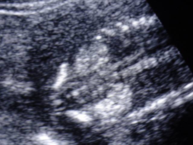
CT Appearance of Acute Appendagitis

OBJECTIVE: Our aim was to describe the spectrum of CT findings in patients with acute epiploic appendagitis and also to evaluate the changes seen with this condition.

MATERIALS AND METHODS: Fifty patients diagnosed with acute epiploic appendagitis seen on contrast-enhanced CT were included in this study. The CT scans of the epiploic appendagitis were evaluated for the presence of colon wall thickening, a focal fatty center, inflammatory changes, location in relationship to the colon, size, and presence or absence of central high density within the fat. In 10 patients, the initial findings were compared with findings of follow-up CT performed between 3 days-21 months after the first CT.

RESULTS: The most common part of colon involved by acute epiploic appendagitis was the sigmoid colon (31/50), and the most common position was anterior to the colonic lumen (41/50). All 50 patients with acute epiploic appendagitis had a central fatty core surrounded by inflammation. Colon wall thickening was present in only two, and a central high-density focus was noted only in 27 of 50 patients. In 86% (43/50) of patients, the fatty central core was between 1.5 and 3.5 cm in length. The changes seen on follow-up CT varied, including increased density with a decrease in the size of the fatty central core, no change, complete resolution of findings, and minimal residual density.

CONCLUSION: On CT, acute epiploic appendagitis has a predictable appearance in terms of location, size, and density. The most common finding on CT is a fat-density oval lesion with surrounding inflammation on the anterior aspect of the sigmoid colon. The changes on CT are not predictable in the 2-week to 6-month window.

AJR Am J Roentgenol. 2004 Nov;183(5):1303-7.

Wednesday, November 3, 2004


Pulmonary angiography showing a Rasmussen's aneurysm of the right lower lobe incidentally discovered in a patient with a previous history of cavitary tuberculosis of the same lobe.

COPYRIGHT (SUMER)

Tuesday, November 2, 2004


phantom tumour-collection of pleural fluid between lobes, i.e. within the major and minor fissures. The radiographic appearance depends on the shape and orientation of the fissure, the volume of fluid, its position within the fissure and the radiographic projection. Interlobar fluid is particularly common in heart failure.

COPYRIGHT (SUMER)

Scurvy
Rare before 6 months of age since the storage of vitamin C in neonate is generally adequate
• Wimberger sign: presence of a sclerotic rim around epiphysis
• White line of frankel:dense zone of provisional calci­fication at the growing metaphysis
• Trumerfeld zone:a lucent zone below white line due to lack of mineralisation
• Pelkan spur:as the area is prone to fractures manifesting at cortical margin
• Osteoporosis
• Subperiosteal haemorrhage.
COPYRIGHT (SUMER)

Tutorials

STRING OF BEADS


PULMONARY EDEMA


BILIARY ATRESIA


MITRAL STENOSIS


MRI IN BLADDER CANCER


RADIOBIOLOGY


COPD X-RAY FINDINGS


WHITAKER TEST


CHYLOTHORAX


PULMONARY INFECTIONS


RADIOSENSTIVITY OF TUMOURS


DYSPHAGIA


ERCP VS MRCP


RADIATION UNITS


SCURVY


Tc99 decay scheme


Monday, November 1, 2004


Langerhans' cell histiocytosis

Lateral radiograph of the skull demonstrating multiple lytic lesions of histiocytosis. The anterior lesion has a "geographical" pattern.


COPYRIGHT (SUMER)

imaging news

Portable CT goes to ICU patients


Portable CT scanners can now provide valuable abdominal diagnostic information for intensive care unit patients without forcing them to leave the unit, according to a study in the September issue of the American Journal of Roentgenology.

The first portable CT scanners were deployed at several large institutions in the late 1990s. Little is known, however, about their performance and use in addition to cranial imaging, which remains portable CT's main application. Unlike cranial CT, an abdominal scan requires moving patients from their beds onto the scanning table. But portable scans acquired at the bedside can reduce well-documented risks associated with transport of ICU patients to other hospital locations.

Dr. Michael M. Maher and colleagues at Massachusetts General Hospital retrospectively reviewed spiral portable abdominal CT scans from 107 patients obtained between June 1999 and December 2000. They compared portable CT's image quality and diagnostic value with that of available stationary CT scans.

Although image quality from portable abdominal CT did not match stationary CT, the researchers found they were able to obtain important diagnostic information without moving patients from the ICU.

Researchers obtained 122 portable and 41 stationary CT scans, with 47 and 15, respectively, enhanced by contrast. Intravenous contrast improved portable CT scan quality. Quality scores for portable CT scans, however, were consistently lower than those for stationary CT, both with and without contrast.

Findings on portable CT confirmed 33 conditions suspected before scanning. Portable scanning also detected evidence of infection in 18 patients and hemorrhage in 16, led to seven laparotomies and six percutaneous drainage procedures, and influenced a change in patient management in 33 cases. Surgery or autopsy results confirmed portable CT findings in 12 of 17 cases.

The spiral portable CT scanning protocol included two 285 to 355-mm-long volumes acquired with 5-mm slice thickness during 70 sec at 120 to 130 kVp, 30 to 40 mAs, and a 1 to 1.5-sec pitch. Patient condition and imaging indication determined IV contrast use. Stationary scanning was performed with single- and four-detector spiral scanners.

Interpretation of portable CT studies must proceed with caution, however. Some portable CT parameters, such as accuracy and negative predictive value, were unknown. While portable scanning is useful in many cases, patients who require valuable diagnostic information should be moved to more sophisticated imaging installations whenever possible, researchers said.


Can MRI replace DMSA in the detection of renal parenchymal defects in children with urinary tract infections?

Background: Renal parenchymal defects may be a consequence of urinary tract infections (UTI) in childhood. MRI is a non-radiation imaging modality compared with DMSA scanning. Objective: To compare DMSA with MRI for the detection of renal parenchymal defects in children presenting for radiological investigation after a first UTI. Materials and methods: Both DMSA and MRI were performed at the same appointment in 37 children (aged 4 months-13 years; mean 4.5 years) with a history of UTI. Both planar and SPECT DMSA were performed. MRI of the kidneys employed axial and coronal T1-, T2- and fat-saturated T1-weighted (T1-W) sequences. Some children had imaging after IV contrast medium. Results: The coronal fat-saturated T1-W sequence was the best sequence and it detected all the findings on MRI. MRI had a sensitivity of 77% and a specificity of 87% for the detection of a scarred kidney using DMSA as the gold standard. MRI diagnosed pyelonephritis in two children that had been interpreted as scarring on DMSA. Conclusions: Renal MRI using a single, coronal, fat-saturated T1-W sequence is a rapid, accurate and minimally invasive technique for the the detection of renal scarring that does not employ ionizing radiation.


Pediatr Radiol. 2004 Oct 14 [Epub ahead of print]

Sunday, October 31, 2004


Sequestrum- A large rod-like sequestrum is observed in the medullary canal owing to chronic osteomyelitis.

COPYRIGHT (SUMER)

Chest radiograph demonstrates decreased pulmonary vascularity, normal cardiac size, concave main pulmonary artery segment and right aortic arch... the boot shaped heart..or cor-en-sabot..

COPYRIGHT (SUMER)

Saturday, October 30, 2004

STRING OF BEADS

string of beads in radiology is a sign which has been described for various pathologies.. it is seen in--


String of beads in radiology-

  • Fibromuscular dysplasia

  • Chr. Pancreatitis (chain of lakes)
  • Small bowel obstruction
  • varicose bronchiectasis


Fibromuscular dysplasia

condition of unknown aetiology that involves the extracranial internal carotid arteries and vertebral arteries. The internal carotid is involved in about 75% of cases and the vertebral in less than 25%. It is characterized by narrowing of the affected vessel with a string of beads appearance , due to focal annular repetitive intimal and medial proliferative changes. Not infrequently an incidental finding, fibromuscular hyperplasia may be a cause of dissection and is associated with an increased incidence of intracranial aneurysms.
COPYRIGHT (SUMER)

Pulmonary edema

Pulmonary oedema


The radiographic changes of hydrostatic oedema are quite characteristic. In the normal adult, the lower lobe pulmonary vessels are larger than the upper lobe vessels due to gravitational forces. As the left-sided pressure increases, the blood is diverted to the upper lobes. This results in "cephalization" with the upper lobe vessels becoming larger than the lower lobe vessels. As left heart pressure increases, fluid enters the peribronchovascular interstitium. As the interstitium becomes oedematous, the interlobular septa become prominent and the markings indistinct.  Pleural effusions are frequent in the more severe stages of left heart failure with a slight predominance to the right.

Thursday, October 28, 2004


Lateral radiograph of a lumbar vertebral body demonstrates the condensation of the bone along the periphery of the expanded vertebral body which has been termed a "picture frame" appearance
classical of pagets disease
COPYRIGHT (SUMER)

Paget's disease
Lateral radiograph of the skull demonstrates a thickened calvarium and fluffy radiodense regions which is referred to as a "cotton-wool" appearance.

COPYRIGHT (SUMER)

Barium swallow in a child showing typical appearances of achalasia. Note the distended oesophagus with food debris, tapered distal end and absence of stomach bubble.


COPYRIGHT (SUMER)

Wednesday, October 27, 2004


sheperd's crook deformity femur-typical of fibrous dysplasia
COPYRIGHT (SUMER)

Tuesday, October 26, 2004


air bronchogram-- a classical sign described by fleishner.. common causes are- pneumonic consolidation, pulmonary edema and hyaline membrane disease
COPYRIGHT (SUMER)

Monday, October 25, 2004


small densely calcified nonfunctioning right kidney, due to longstanding tuberculosis. the so called autonephrectomy/PUTTY KIDNEY


COPYRIGHT (SUMER)

IMAGING IN BILIARY ATRESIA

biliary atresia

a condition in which there is aplasia or obliteration of some or all of the extrahepatic biliary tree: the gallbladder may or may not be involved. It presents in the neonate or young infant with clinical findings of obstructive jaundice and conjugated hyperbilirubinaemia. If undiagnosed or unrelieved progressive biliary cirrhosis will develop. The prognosis is inversely related to the age at which surgery is undertaken and the degree of liver damage. The aetiology is unknown but some consider it to be due to an inflammatory process which leads to progressive obliteration of the bile ducts in the perinatal period.


At ultrasound examination, which should be done following a 4-hour fast, before the onset of cirrhosis, the liver is normal. There is no intrahepatic bile duct dilatation. The gallbladder is not usually seen but if identified it may be spherical. The extrahepatic biliary tree is not seen. A normal ultrasound examination does not exclude the diagnosis. Hepatobiliary scintigraphy using a Tc99m-labelled iminodiacetic acid product (e.g. HIDA) typically demonstrates good hepatic uptake of tracer, with progressive accumulation within the liver but no evidence of excretion into the extrahepatic bile ducts or into bowel. Premedication with phenobarbitone for 5 days prior to the study is recommended to ensure maximal hepatic enzyme function should there be some impairment of liver function. The absence of bowel activity at 24 hours is generally taken to be an indication for liver biopsy and/or operative cholangiography. Findings in neonatal hepatitis include poor hepatocellular uptake of tracer, delayed excretion into bowel and occasionally non-excretion as in biliary atresia.


Treatment is surgical and involves anastomosing an intrahepatic bile duct (usually in segment III) to a loop of jejunum to bypass the extrahepatic obstruction (Kasai procedure). Complications include sepsis and anastamotic stricture. The procedure should be performed as early as possible after diagnosis to avoid complications related to cirrhosis and portal hypertension.


the classic waterlily sign of hydatid!!!!!
COPYRIGHT (SUMER)

History of X-rays

In the latter half of the year 1895, a German scientist called Roentgen was working in his laboratory at the Physical Institute of the University of Wurzburg, Germany, experimenting with a type of discharge tube called Crooke's tube. The tube displayed a fluorescent glow when a high voltage current was passed through it. When he shielded the tube with heavy black cardboard, he found that a greenish fluorescent light could be seen on a fluorescent screen kept some 9 feet away. Roentgen concluded that a new type of ray was emitted from the tube that could pass through the black covering. The rays could pass through most substances, including the soft tissues of the body, but left the bones and most metals visible. One of his earliest photographic plates from his experiments was that of a film of his wife, Bertha's hand with a ring. Roentgen named the invisible radiations as X-rays (or unknown rays).

indian conference calender

CMEs and Conferences In India : Information about CMEs and conferences in India

2-4-2005 to 3-4-2005
22nd Karnataka state level conference of I.R.I.A By-D.K chapter of Karnataka state Branch of IRIA At-Dr.T.M.A Pai Convention centre Mangalore Contact :Dr.Raghavendra Bhat. K Address : Balmatta scan Centre.Balmatta Mangalore PH: 0824-2443720,9845071520 Email : rbhatk@sancharnet.in
------------------------------------------------------------------- --------------------------------------------
8-4-2005 to 10-4-2005
Clinical Course and Hands - on training for Proton MRS of Brain. By-MRI centre, Dr. Balabhai Nanavati Hospital, Mumbai in collaboration with GE healthcare, India At-Dr. Balabhai Nanavati Hospital, S.V. Road, Vile Parle (w), Mumbai - 56 Contact :Dr. Sona Pungavkar,Mobile No. 098202855565Address : Dr. Balabhai Nanavati Hospital, S.V. Road, Vile Parle (w), Mumbai - 56 Ph : 098202855565
Email : drsonap@yahoo.co.in Website www.nanavatihospital.org
--------------------------------------------------------------------------------------------------------------
9-4-2005 to 10-4-2005
North Zone Radiology Meet 2005 By-IRIA, Punjab & Chd branch At-Govt. Medical College, Patiala Organiser : Dr. Manoj Mathur Contact : manojnidhi66@rediffmail.com Address : 2014 Lal Bagh Street,Patiala Ph : 0175-2212246, 094173-29926 Email : manojnidhi66@rediffmail.com
----------------------------------------------------------------------------------------------------------
30-4-2005 to 1-5-2005
3rd Chest Radiology Review Course By-REF,IndiaAt-MLT, KEM Hospital, Mumbai Address : Bhaveshwar Vihar, 383 Sardar V. P. Rd Ph: 022-2388-4015 Fax: 022-2382-9595 Email : info@refindia.net Website www.refindia.net
--------------------------------------------------------------------------------------------------------------
23-10-2005 to 25-10-2005
ISVIR - 8th Annual Conference At-Pune
Contact : Wg Cdr Hirdesh Sahni Address : Associate Professor , Dept of Radiodiagnosis, AFMC, Solapur Rd, Pune 411040 Ph: 020-26306061, 09370144728 Email : isvir2005@rediffmail.com Website www.isvir.org
----------------------------------------------------------------------------------------------------------

Sunday, October 24, 2004


hydatid cyst lung.. a classical CXR appearance although this is not exactly water lily appearance but it is quite specific.. always remember hydatid in lung almost never calcifies.


COPYRIGHT (SUMER)

Saturday, October 23, 2004

MITRAL STENOSIS-RADIOLOGICAL FINDINGS

Mitral Stenosis:


Clinical:The normal area of the mitral valve is 4 to 6 cm2. Severe stenosis is associated with areas less than 0.8 cm2, and most valves are replaced when their area becomes less than 1.1 cm2. Mitral stenosis results in increased resistance to emptying of the left atrium. This produces a reduced left ventricular output and an increase in pulmonary venous pressure (a valveless system, thus LA pressures are transmitted directly to the vessels). Eventually this increased pressure is back transmitted to the pulmonary arteries. The pulmonary arteries undergo medial hypertrophy and intimal sclerosis in response, and pulmonary arterial hypertension results. Ultimately, the right ventricle hypertrophies, dilates, and then fails. Clinically there is a diastolic murmur and resting tachycardia as the heart attempts to supply more blood systemically. Rarely, patients with mitral stenosis can present with diffuse alveolar hemorrhage [2]. Another rare, late sequella of mitral stenosis is parenchymal ossification [2]. In approximately 0.6% of cases of mitral stenosis, a coexisting ASD may relieve the left atrial hypertension and promote the formation of a left to right shunt. This combination of findings is known as Lutembacher syndrome [2].

The two major factors influencing prognosis in mitral stenosis are the presence of pulmonary hypertension (triples operative mortality) and the presence of symptoms [3]. Once more than mild symptoms develop, the prognosis for medical treatment decreases [3].

Etiologies of mitral stenosis include rheumatic heart disease (most commonly), congenital mitral stenosis, or an obstructing lesion such as a left atrial myxoma.

X-ray:

CXR: The left atrial appendage is the only portion of the left atrium that forms part of the left border of the heart. On PA radiographs it occupies the portion of the left heart border between the main pulmonary artery segment and the superior portion of the left ventricular contour. When left atrial pressure and volume are normal, this segment of the left heart border is concave. Early or mild enlargement of the left atrium may be detected as enlargement of the left atrial appendage with straightening of this segment of the left heart border. With continued enlargement, this segment will become convex. Another finding of left atrial enlargement include a "double density" in the mid-portion of the cardiac silhouette on the frontal view. A line from the mid-point of the right border of the double density to the midpoint of the border of the left mainstem bronchus should measure less than 7.5 cm (7.0 cm in females). A normal left atrium should also lie anterior to a line drawn down the center of the trachea on the lateral, non-rotated view. Other findings which can suggest LA enlargement include posterior esophageal displacement on barium swallow, elevation of the left mainstem bronchus, and straightening of the left heart border due to enlargement of the left atrial appendage. In cases of long-standing stenosis, the LA wall may calcify. Mitral valve calcification is only seen 10% of cases (Note: Calcification of the mitral valve annulus does not indicate mitral stenosis). Pulmonary venous congestion can be seen as the stenosis progresses. Since the left ventricle is unaffected by mitral stenosis it will remain normal [2]. Later there is pulmonary arterial hypertension and right ventricular enlargement.

MRI: On MRI other findings of mitral stenosis include a mild increased signal intensity in the lungs on spin echo images due to pulmonary venous hypertension and interstitial edema. Cine gradient images can be used to demonstrate turbulent flow across the mitral valve which appears as a fan-shaped signal void in the LV below the valve during diastole.


Friday, October 22, 2004

on line book purchasing

NEW NEW NEW...... NOW AVAILABLE

All India PG entrance exam 2005 fully solved by Dr Sumer K Sethi and Dr Sidharth K Sethi is now available in the market!!!
this is only authentic solution to aipg 2005 available till now, it has explanatory answer from latest editions of standard textbooks with extensive online support at
www.sumerdoc.blogspot.com

AIIMS NOV 2004 FULLY SOLVED AND EXPLAINED

aiims nov 2004 fully solved and explained by sumer sethi is availlable in the market...highlights of the book are authentic references, diagrams and flowcharts, extensive coverage of topics so that all repeat questions are tackled...and first book of its kind to give extensive online support with an online discussion forum support by the author.. also by the same author "review of radiology" solving quite a few questions in this aiims!! and may 2004 aiims fully solved peepee publishers

to order any book of peepee publishers for pg entrance preparation- including AIPG JAN 2005(Rs165/-), NOV 2004 AIIMS (Rs150/-),MAY 2004 AIIMS (Rs 95/-) solved by me and Review Of Radiology (Rs100/-) a must for all PG aspirants for radiology

simply fill in ur details including name, email address and phone number and mailing address.. write or mail to

peepee160@rediffmail.com

and address-

Peepee publishers and distributors (P) ltd

7/31 ansari road, darya ganj,

post box no-7243

new delhi 110002

india

SEND A CHEQUE/ D/D TO THE ABOVE ADDRESS

SPECIAL OFFER-NO POSTAGE WUD BE REQUIRED FOR ORDERS IN INDIA

----------------------------------------------------------------------------------------------------------

ALSO ON--
MY BOOKS AVAILABLE ONLINE-- NO BOOK SHOPS REQUIRED

my book "review of radiology" if it is not available in your city or if u ve any problem purchasing the book check out the link-


http://www.mcqsonline.com/edoctor/radiology_review.htm

also if u want MAY 2004 solved paper by sumer sethi extensively researched solutions to may 2004 aiims click here--


http://www.mcqsonline.com/edoctor/aiims_2004.htm

also available at
http://www.geocities.com/grajesh70/bookstore.htm

FOR NOV AIIMS TO ORDER ONLINE--

http://www.edoctor.mcqsonline.com/aiims_nov_2004.htm

TO ORDER ASHWIN UDAYAR'S MAHE GUIDE WITH 2000-2004 PAPERS

www.mcqsonline.com/edoctor/mahe_2004.htm


cranial USG showing bilateral subdural effusion.. cranial USG is a useful technique for infants to look for hydrocephalus, subdural effusion and intracranial bleeds
COPYRIGHT (SUMER)

choledocolithiasis-the classical meniscus sign at the distal end of CBD
COPYRIGHT (SUMER)

Wednesday, October 20, 2004

MR imaging bladder

abstract

Magnetic resonance imaging (MRI) of the bladder is most frequently used for staging of a known bladder tumor and follow-up of a treated cancer patient. MRI is well suited for bladder evaluations, since it is a noninvasive examination that allows multiplanar visualization of the entire pelvis and offers superior soft-tissue contrast.


Bladder cancer is a common tumor of the urinary tract, accounting for 6% to 8% of male malignancies and 2% to 3% of female malignancies.1 The most common histologic cell type is transitional cell carcinoma, accounting for 90% of all primary tumors of the urinary bladder. The remaining 5% to 10% of malignant bladder tumors are nontransitional and consist of squamous cell carcinomas, adenocarcinomas, small cell carcinomas, and, rarely, sarcomas. Cross-sectional imaging aids in determining tumor stage, including detection of local extension of the tumor, lymph node involvement, and distant metastasis.2 Presently, magnetic resonance (MR) imaging is the modality of choice in imaging of urinary bladder neoplasms, and staging accuracy of MR imaging in bladder cancer ranges from 73% to 96%. These values are 10% to 33% higher than those obtained with computed tomography (CT).3 Because the most common disease of the urinary bladder that requires further imaging is bladder cancer,4 the most frequent indications for MR imaging of the bladder are staging of a known bladder tumor and follow-up of a treated cancer patient. The excellent soft-tissue contrast and multiplanar capability of MR imaging improve the evaluation of the location and extent of bladder tumors.5


Technique


Currently, a 1.5T MR scanner with a phased-array pelvic coil is commonly used for imaging of the urinary bladder. The typical urinary bladder protocol includes T1-weighted spin-echo images (repetition time [TR]: 400 to 550 msec; echo time [TE]: minimum) obtained in the axial plane and T2-weighted fast spin-echo (TR: 4000 to 5500 msec; TE: 80 to 120 msec) images also obtained in the axial plane. Subsequently, fast multiplanar spoiled gradient-echo images with fat suppression (TR: 180 to 300 msec; TE:1.7 to 4.2 msec) are obtained in the axial plane before, at 20 seconds (arterial phase), and at 70 to 115 seconds (venous phase) after gadopentate dimeglumine injection (0.1 mmol/kg). Sagittal or coronal T2-weighted images may be obtained, if the anteroposterior or inferosuperior extent of disease needs to be evaluated. Sagittal and coronal gadolinium-enhanced images are also recommended for the evaluation of the bladder tumors, when the tumor is located in the base or the dome of the bladder. Typical parameters for optimal bladder imaging are: 20- to 30-cm field-of-view (FOV), 6-mm slice thickness, and 2-mm intersection gap. Ideally, the urinary bladder should be moderately distended during imaging. The low distension may cause misevaluation of the bladder wall thickening, and overdistension may obscure small tumors.


Bladder cancer


Bladder cancer is more common in men (male/female: 3:1) than in women. It is the fourth most common cancer in men, after prostate, lung, and colorectal cancer, and it accounts for 10% of all cancer cases in men. In women, it is the eighth most common cause of cancer and accounts for 4% of all cancer cases in women.6 It is generally a disease of the elderly, and its incidence peaks at the sixth to seventh decade.7


Several factors predispose patients to bladder cancer. Cigarette smokers are 2 to 6 times more likely to develop urothelial cancer than those who do not smoke.8,9 Cystitis and chronic urinary tract infection are predisposing factors to bladder cancer, and 7% of bladder tumors are found to be associated with diverticuli (Figure 1). Strong association with aniline dyes, aromatic amines, diesel fumes, and long-term use of analgesic phenacetin has been reported in the development of bladder cancer.1


Pathologically, 90% of the bladder tumors are epithelial, and 90% of the epithelial tumors are transitional cell carcinomas. At the time of presentation, 30% of patients have multifocal disease, and there may be widespread areas of metaplasia and carcinoma in situ.1 The remaining 10% of epithelial tumors include squamous cell carcinomas, adenocarcinomas, small cell carcinomas, and, rarely, sarcomas.2 Leiomyosarcoma, rhabdomyosarcoma (Figure 2), phaeochromocytoma, and hamartomatous malformations are among the nonepithelial tumors.1 Gross or microscopic hematuria is the most common clinical presentation, followed by symptoms related to associated urinary infection.10


Staging and management of bladder cancer


Local extension of the tumor (T), lymph node involvement (N), and distant metastasis (M) are the indicators used in staging. Table 1 presents the bladder cancer staging used.11 Treatment options and prognosis depend on the clinical stage of the bladder tumor. It is critically important to differentiate between superficial and invasive disease.1 Superficial tumors are treated with local endoscopic resection, with or without adjuvant intravesical instillations of chemotherapeutic agents; while invasive tumors are treated by curative cystectomy and palliative chemo- and/or radiation therapy.12


Management of bladder tumors starts with clinical staging, including cystoscopic examination of the organ. This method allows for immediate biopsy and can distinguish superficial from invasive tumors, but it is not capable of detecting extravesical disease. This distinction is important because patients with non– organ-confined disease have higher recurrence rates and lower survival rates.13


CT has been a valuable tool for the evaluation of bladder tumors, but MR imaging has been shown to be superior in the detection of superficial and multiple tumors, and in staging accuracy in detecting extravesical tumor extension and surrounding organ invasion, especially with dynamic contrast administration.2,14-20 Accuracy of MR imaging on a stage-by-stage basis has been reported to be between 64% and 85%.16,18,19,21,22 Staging accuracy of MR imaging in differentiating superficially versus deeply muscle-invasive tumors, and organ-confined versus non–organ-con-fined tumors is between 85% and 95%.15,17,21,23


Restaging after treatment is particularly challenging. Intravesical therapy with chemotherapeutic agents is used to treat low-stage tumors. Intravesical therapy may cause bright submucosal enhancement after administration of gadolinium. Previoustransurethral resection or biopsy of the tumor may cause inflammation and edema, and this may result in overstaging. Using MR imaging, it is not possible to discriminate by signal characteristics alone between edema resulting from treatment and tumor recurrence, but the presence of a mass with an enhancement pattern typical of a tumor may enable the diagnosis of recurrence.


Morphologic features


Tumors can arise anywhere in the bladder, but they are most commonly located in the lateral wall. When a tumor is located around the ureteral orifices, it may produce partial or complete blockage of one or both ureters, resulting in hydroureter and hydronephrosis.10 Tumors manifest a variety of patterns of growth, including papillary, sessile, infiltrating, nodular, mixed, and flat intraepithelial growth. Because the bladder does not have a distinct basement membrane, it is difficult to detect the invasion of the lamina propria by imaging .24 However, the detrussor muscle can be well evaluated with MR imaging; therefore, the depth of muscle invasion can be assessed. The fat tissue around the bladder can be seen clearly on MR imaging, and the interface between the bladder wall and the surrounding fat can be evaluated for extravesical tumor spread.


MR imaging features


Urinary carcinomas have intermediate signal intensity, equal to that of muscle on the T1-weighted images. T1-weighted images are used to assess perivesical fat invasion and lymph node involvement. T2-weighted images aid in determining the depth of tumor infiltration into the bladder wall. On T2-weighted images, urine has high signal intensity, and the bladder wall appears hypointense. Bladder tumors have the same, or slightly higher, signal intensity as the bladder wall on T2-weighted images. An intact, low-signal-intensity muscle layer at the base of the tumor is classified as stage Ta or T1 (Figure 3); a disrupted low-signal-intensity muscle layer without infiltration of perivesical fat is classified as stage T2b (Figure 4).25


Urinary bladder carcinomas and their metastases develop neovascularization26; therefore, these tumors enhance earlier than the normal bladder wall.3 In the arterial phase of contrast enhancement, bladder tumors enhance more than the muscle of the bladder (Figure 5). Superficial tumors (those without muscle invasion, stage T1 and lower) may be differentiated from muscle-invasive tumors on contrast-enhanced images. On contrast-enhanced studies, a lesion with an irregular, shaggy outer border, and streaky areas of the signal intensity of the tumor in the perivesical fat is classified as stage T3b (Figure 6). A tumor extending into an adjacent organ or abdominal and pelvic sidewall with the same signal intensity as the primary tumor is classified as stage T4a or T4b, respectively (Figure 7).25With the current imaging protocols using 1.5T MR scanners, MR imaging cannot differentiate stage Ta tumors from stage T1; and differentiation of stage T2a tumors (superficial muscle invasion) from stage T2b (deep muscle invasion) is problematic but sometimes can be better delineated on contrast-enhanced studies.2


Based on MR imaging features, it is not possible to differentiate between different histologic cell types of bladder tumors, but few clues may lead to the differentiation of transitional from non–transitional-cell carcinomas. Non–transitional-cell carcinomas are aggressive tumors that usually extend beyond the bladder wall at the time of initial diagnosis, whereas two thirds of transitional cell carcinomas are superficial. Non–transitional-cell carcinomas tend to cause marked bladder wall thickening and tend to be larger than transitional cell carcinomas.27 The non–transitional-cell carcinomas of the bladder include: squamous cell carcinoma, adenocarcinoma, small cell carcinoma, and carcinosarcoma.


Squamous cell carcinoma


Squamous cell carcinoma is the most common non–transitional-cell bladder tumor, accounting for 3% to 7% of all bladder tumors in the United States. Approximately 80% of squamous cell carcinomas in Egypt are associated with chronic infection caused by schistosomiasis (bilharziasis).24The disease is relatively more common in women, unlike transitional cell carcinoma, which is more common in men. The reported female-to-male ratio varies from 1.25:1 to 1.8:1.28 In contrast to transitional cell carcinomas, squamous cell carcinomas are often widespread and involve areas other than the base of the bladder. Most squamous cell carcinomas are solitary and large at the time of detection, with invasion of the muscular wall reported in >80% of patients (Figure 8). Metastases have been identified in at least 10% of patients at the time of diagnosis. Interestingly, metastases from squamous cell carcinomas of the urinary bladder often occur at sites other than the regional lymph nodes. Common metastatic sites include bone, lung, and bowel.29


Adenocarcinoma


Adenocarcinoma of the urinary bladder is uncommon, accounting for 0.5% to 2% of all bladder malignancies.30 Adenocarcinomas are classified into 3 groups: primary, urachal, and metastatic.24 Like squa mous cell carcinomas, many adenocarcinomas probably occur in reaction to long-term mucosal irritation. Cystitis glandularis, bladder exstrophy, and urachal remnants are also associated with adenocarcinoma of the bladder. Cystitis glandularis has been observed in approximately 50% of adenocarcinoma cases located at the bladder base3,11,24,28,29; however, most au thors do not accept this entity as a predisposing factor for urothelial adenocarcinoma. Adenocarcinoma is observed in <10% of patients with bladder exstrophy, and >80% of urachal neoplasms are adenocarcinomas.29 Signet cell adenocarcinomas characteristically produce linitis plastica of the bladder (Figure 9).


Small cell carcinoma


Small cell carcinoma of the bladder has also been called undifferentiated and poorly differentiated carcinoma. Most small cell carcinomas occur as a component of mixed carcinomas. Age, sex, and symptoms are comparable to those of transitional cell carcinomas. Cystoscopically, small cell carcinomas tend to be polypoid or nodular and often appear as ulcerated masses that cannot be distinguished from other high-grade bladder cancers (Figure 10). Metastatic spread occurs rapidly, and the most frequent sites are the regional lymph nodes, bones, and peritoneal cavity.29


Carcinosarcoma


Carcinosarcoma of the urinary bladder is a rare neoplasm; approximately 73 cases have been reported. They are most frequently observed in the female genital tract.31 Neither the etiology nor the pathogenesis of carcinosarcomas is currently known. Unlike transitional cell carcinomas, carcinosarcomas are highly aggressive tumors, with high recurrence rates and a poor prognosis. These tumors can present as large single tumors or multiple small- to medium-sized tumors. The tumors may be polypoid or sessile and may arise from anywhere in the bladder. Location, size, shape, and multiplicity do not help to differentiate carcinosarcomas from transitional cell carcinomas. Dynamic gadolinium-enhanced imaging in carcinosarcomas does not show early strong arterial enhancement, a feature differentiating this tumor from a transitional cell carcinoma. On T2-weighted imaging, the signal intensity of carcinosarcomas can be heterogeneous.32


Conclusion


MR imaging is a noninvasive examination that allows multiplanar visualization of the entire pelvis and offers superior soft-tissue contrast. It is a useful modality for the assessment of tumors of the urinary bladder. Imaging with gadolinium enhancement allows evaluation of the bladder tumor extent and adjacent organ involvement. MR imaging thus plays a critical role in improving staging accuracy, determining patient management, and assessing response to therapy.


Sunday, October 17, 2004


normal barium meal follow through jejunum has valvulae connvinates or feathery appearance
COPYRIGHT (SUMER)

"pneumatocele" a characterstic of staphylococcal pneumonia
COPYRIGHT (SUMER)

Thursday, October 14, 2004

visit my message board

Visit My Message Board


message board feature has been added to this site to facilitate posting of individual queries and viewpoints. please feel free to express your opinion on it....

"widened intercondylar notch knee" classical feature of hemophilic arthopathy
COPYRIGHT (SUMER)

Wednesday, October 13, 2004

Nonvisualization of Appendix on CT Linked With Low Rate of Appendicitis

Nonvisualization of Appendix on CT Linked With Low Rate of Appendicitis


Nonvisualization of the appendix on helical computed tomography (CT) examination is associated with low incidence of acute appendicitis in the absence of secondary inflammatory changes, according to the results of a retrospective study published in the October issue of the American Journal of Roentgenology. Appendicitis may be safely ruled out given the visualization of more than a scant amount of pericecal fat.

"CT has become part of the standard of care in managing patients with suspected acute appendicitis," writes Paul Nikolaidis, MD, from the Department of Radiology at Northwestern University Feinberg School of Medicine in Chicago, Illinois, and colleagues. "Sometimes, however, the appendix is not visualized on CT examination despite the use of optimal imaging parameters."

To assess the significance of nonvisualization of the appendix in the absence of secondary inflammatory changes, the investigators retrospectively reviewed CTs taken to rule out appendicitis in 366 consecutive patients presenting with symptoms of lower abdominal or right lower quadrant pain.

Original CT reports included 56 CTs (15%) in which the original reviewer was unable to visualize the appendix in the absence of secondary inflammatory changes, including abscess formation, localized perforation, periappendiceal fat stranding, or appendicolith.

The investigators were able to visualize appendices in 10 of these CTs upon review, yielding 46 CTs (13%) in which nonvisualization of the appendix was agreed upon. Pericecal fat was evaluated on a scale of 0 (scant, n = 8), 1 (n = 20), and 2 (abundant, n = 28).

In 12 patients (26%), CT indicated gastrointestinal (n = 8) and genitourinary (n = 4) symptom sources. Of 34 remaining patients, 11 (24%) were diagnosed through further imaging or clinical evaluation. One patient (2%) with a scant amount of pericecal fat (score = 0) was diagnosed with acute appendicitis that was confirmed by surgical pathology.

"The amount of fat surrounding the cecum influences our ability to visualize the appendix and therefore more confidently exclude the possibility of acute appendicitis," the authors write, pointing out the scant amount of pericecal fat (score of 0) in the patient with the missed diagnosis.

Study limitations include its retrospective nature and the lack of follow-up information for a relatively large number of patients (50%).

"In the absence of a distinctly visualized appendix and secondary inflammatory changes, the incidence of acute appendicitis is low (2%)," the authors conclude, adding that acute appendicitis may be safely excluded with CT visualization of more than a scant amount of pericecal fat.


Am J Roentgenol. 2004;183:889-892


OSTEOSARCOMA-"SUNRAY APPEARANCE" Posted by Hello

HEMOLYTIC ANEMIA Posted by Hello

posterior urethral valve Posted by Hello