Tuesday, June 28, 2005

Useful article-Differentiating Malignant from Benign Common Bile Duct Stricture with Multiphasic Helical CT.

Choi SH, Han JK, Lee JM, Lee KH, Kim SH, Lee JY, Choi BI.
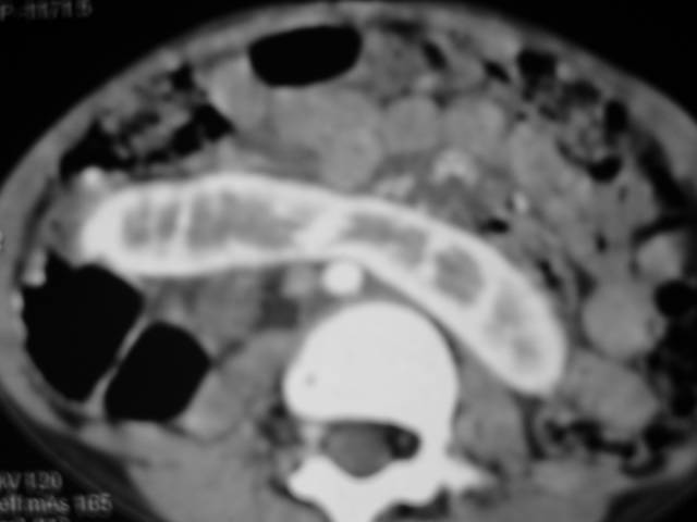
PURPOSE: To evaluate retrospectively the use of multiphasic helical computed tomography (CT) to differentiate malignant and benign common bile duct (CBD) strictures in patients with only a focal CBD stricture and to determine predictors for this differentiation.
RESULTS: Malignant strictures were longer (17.9 mm +/- 6.6 [+/- standard deviation]) than benign strictures (8.9 mm +/- 6.8) (P < .0001), and upstream CBD diameters were larger in malignant cases (22.0 mm +/- 5.4) than in benign cases (17.8 mm +/- 4.6) (P = .033). The involved wall thickness was more than 1.5 mm in 26 malignant cases and three benign cases (P < .0001). During both hepatic arterial and portal venous phases, greater enhancement than that in the normal CBD were more frequently observed in malignant cases (in 27 and 30 patients for hepatic arterial and portal venous phase scans, respectively) than in benign cases (in two and three patients, respectively) (P < .0001). Results of multivariable stepwise logistic regression analysis showed that hyperenhancement of the involved CBD during the portal venous phase was the only variable that could be used to independently differentiate malignant from benign strictures.
CONCLUSION: Hyperenhancement of the involved CBD during the portal venous phase is the main factor distinguishing malignant from benign CBD strictures.
Radiology. 2005 Jun 13; [Epub ahead of print]

Thursday, June 23, 2005

Medical Gadget Information on www.indianradiology.com, the most happening radiology site in India

Image hosted by Photobucket.com
‘Elektron Co.’ is a Russian company in medical radiology business.It has dealt with R&D and manufacture of X-Ray machines and OEM components since 1989. The basic direction of its R&D is digital imaging for medical purposes.‘Elektron Co.’ is the leader in development and production of X-Ray medical equipment in Russia. The company has first-class production facilities and scientific resources, which provide for high efficiency of hi-tech manufacture in accordance with world quality requirements. Its staff numbers more than 300 employees. Among them are opticians, mechanics, experts on electronics and programming, highly skilled specialists, which are able to implement any engineering design.In 2004 ‘Elektron Co.’ has completed construction of its new research and production facilities in St. Petersburg, Russia. The company has implemented and become registered to the ISO 9001:2000 Quality Management System.‘Elektron Co.’ offers for purchase the following equipment: FC-01 - digital radiography system for chest diagnostics and screening; DIRA - high-resolution multi-function digital radiography system; KRT and RDK - multi-function radiology systems; RTC - mobile C-Arm system. All the systems have been certified by Russian Ministry of Health. The equipment of ‘Elektron Co.’ is competitive with its opponents originated from well known world leading companies.High competitiveness of the company equipment is proved by its regular successful tender bids not in Russia only but also overseas (EBRD, NATO, etc.). ‘Elektron Co.’ successfully cooperates with such companies as Thales, IAE, Mecall and many others.Now the company supplies its equipment to markets of Russia, Western Europe and China. ‘Elektron Co.’ is planning to expand and enter markets of America and Africa and also to increase its share of the current markets.For further information please contact export@elektron.ru.

This is not a commercial advertisement. Owners of the site (Sumer’s Radiology Site) are not in anyways responsible or guarantee the products or the information provided. And are in no ways related to the products shown in the post. This is just a beginning of the effort to familiarize the readers of this online radiology magazine with new products useful for Radiologists or Physicians available in the market.

Journal watch-MRI of the cardiomyopathies.

Di Cesare E.
Authors examined the potentialities of Magnetic resonance imaging (MRI) in the evaluation of the main cardiomyopathies: hypertrophic, dilated, restrictive and arrhythmogenic right ventricular. The hypertrophic cardiomyopathy is generally adequately investigated by echocardiography, that well defines the myocardial thickening and the obstruction of the left ventricular output. However, by echocardiography we still have difficulties in the evaluation of the apex of the left ventricle and the right ventricle involvement. MRI provides a complete evaluation of the heart with a clear evidence also of the echocardiographic dark zones by means of a clear evidence of the apex of the right ventricle. The dilated form is also well investigated by MRI that provides a clear evaluation of the volumes, mass and ejection fraction by means of the 3D analysis including conditions of the ventricular remodelling. Moreover, this technique helps in the differential diagnosis of acute myocarditis. In the acute phase of myocarditis (first 2 weeks), in fact, the myocardium produces high signal intensity on the T2 weighted sequences due to the presence of oedema. The third form of cardiomyopathy is the restrictive one, characterised by reduced diastolic filling and diastolic volume, normality of the systolic function and parietal thickness, interstitial fibrosis and enlargement of both atria. The mean potentiality of MRI is related to the differential diagnosis with constrictive pericarditis. Only in the former, the pericardium appears irregularly thickened with areas exceeding 4 mm of pericardial thickness. Finally, the right ventricular arrhythmogenic cardiomyopathy represents the main indication to MRI evaluation. With this imaging modality we are can obtain a clear morpho-functional evaluation of the right ventricle and distinguish the intramyocardial adipose substitution characterised by areas of high signal in the myocardium.
Eur J Radiol. 2001 Jun;38(3):179-84.

Wednesday, June 22, 2005

Answer to the image quiz

Answer-Congenital Diaphragmatic Hernia
Winner-Jen SN (email sumerdoc-AT-yahoo-dot-com with your mailing address)
About Diaphragmatic Hernia-
The topic of congenital diaphragmatic hernia (CDH) frequently appears in the medical literature since its first description in the early 18th century. Initial theories about the pathophysiology of this condition centered on the presence of the herniated viscera within the chest and the need for its prompt removal. In 1946, Gross reported the first successful repair of a neonatal diaphragmatic hernia in the first 24 hours of life. The medical literature for the next decade addressed CDH as a surgical problem and discussed various technical aspects of surgical repair, including techniques required to close large defects. In the 1960s, however, Areechon and Reid observed that the high mortality rate of CDH was related to the degree of pulmonary hypoplasia at birth. Over the past 20 years, pulmonary hypertension and pulmonary hypoplasia have been recognized as the 2 cornerstones of the pathophysiology of CDH. In recent years, evidence suggests that dysfunction of the surfactant system as well as cardiac maldevelopment may further complicate the pathophysiology of CDH.
The 3 basic types of CDH are the posterolateral Bochdalek hernia (occurring at approximately 6 weeks' gestation), the anterior Morgagni hernia, and the hiatus hernia. The left-sided Bochdalek hernia occurs in approximately 90% of cases. Left-sided hernias allow herniation of both small and large bowel as well as intra-abdominal solid organs into the thoracic cavity. In right-sided hernias, only the liver and a portion of the large bowel tend to herniate. Bilateral hernias are uncommon and usually fatal.
Further Reading- Article on eMedicine

Tuesday, June 14, 2005

Interesting abstract-Placental volume measured by three-dimensional ultrasound at 11 to 13 + 6 weeks of gestation: relation to chromosomal defects.

Wegrzyn P, Faro C, Falcon O, Fabio C, Peralta CF, Nicolaides KH.
OBJECTIVE: To determine the potential value of measuring the placental volume at 11 to 13 + 6 weeks of gestation in screening for chromosomal defects.
METHODS: The placental volume was measured using three-dimensional ultrasound in 500 consecutive singleton pregnancies immediately before chorionic villus sampling for fetal karyotyping at 11 to 13 + 6 (median, 12) weeks of gestation.
RESULTS: The fetal karyotype was normal in 417 pregnancies and abnormal in 83. In the chromosomally normal group the mean placental volume increased significantly with gestation from a mean of 51 mL (5th and 95th centiles: 31.2 and 82.4 mL) at 11 weeks to 91 mL (5th and 95th centiles: 55.7 and 147.2 mL) at 13 + 6 weeks. In the chromosomally abnormal group the mean placental volume for gestational age was not significantly different from normal in trisomy 21 and Turner syndrome, but it was smaller in trisomies 13 and 18.
CONCLUSIONS: The measurement of the placental volume at 11 to 13 + 6 weeks of gestation is unlikely to be a useful predictor of the major chromosomal defects. In trisomies 13 and 18 the small placental volume may be due to early-onset fetal growth restriction, which could be the consequence of impaired placental function.

Monday, June 13, 2005

Image Quiz-Pediatrics


Image Quiz

This neonate presented to the neonatologist in the labour room with respiratory distress at birth. CXR was done. Can you make the diagnosis?

First person to make the correct diagnosis recieves a complimentary copy of "Review Of Radiology" by me.
COPYRIGHT (SUMER)

Saturday, June 11, 2005

Journal watch-How safe is magnetic resonance imaging in patients with contraceptive implants?

Muhler M, Taupitz M.
When patients with an implanted contraceptive device undergo MRI, it must be ensured that the examination involves no risk to the patient (MR safety) and that the diagnosis is not affected by artifacts or the function of the device compromised (MR compatibility). Two basic types of intrauterine devices can be distinguished: the metal-containing/metal-free intrauterine device (IUD) and the hormone-containing implant, the fully metal-free intrauterine system (IUS), as well as the ESSURE insert made of stainless steel, which has been approved for use in Europe since February 2001.The metal-containing and metal-free IUDs and ESSURE are MRI compatible up to a magnetic field strength of 1.5 T. They do not interact in any relevant way with the external magnetic or high-frequency field and the temperature increase is within the physiologic range. The implants merely produce a local signal void with a shape that depends on their orientation relative to the magnetic field lines. At 3 T, only the metal-free IUD and the IUS are MRI safe in terms of the material used. In contrast, metal-containing IUDs and the ESSURE have not yet been fully evaluated in the 3 T field, which is why they represent a contraindication to MRI. No data are available on the MRI compatibility at 3 T for any of these devices.

Tuesday, June 7, 2005

Image Case-Horse shoe kidney


Contrast enhanced CT image showing fusion of the lower poles of the kidneys-Horse shoe kidney
COPYRIGHT (SUMER)

Saturday, June 4, 2005

Interesting abstract-Renal sonographic findings of type I glycogen storage disease in infancy and early childhood.

Lin CC, Tsai JD, Lin SP, Lee HC.
Background: Type I glycogen storage disease (GSD-I) is an inherited disorder affecting glycogenolysis and gluconeogenesis. The characteristic manifestations are hepatomegaly, hypoglycemia, hyperlacticacidemia, hyperuricemia, and hyperlipidemia. Renal disease is regarded as a long-term complication and is reported mainly in older patients.
Objective: We report the renal manifestations and renal ultrasonographic findings of GSD-I in infancy and early childhood in order to assess the role of renal sonography in the diagnosis of GSD-I.
Materials and methods: We retrospectively reviewed our hospital's database for patients with GSD-I from January 1993 to September 2004. The records of five patients were reviewed for this study. These five patients were diagnosed when they were younger than 3 years old. Data extracted from the charts included the initial extrarenal and renal manifestations, laboratory data, and imaging studies. We analyzed the indications for, and results of, renal sonography.
Results: In addition to the clinical presentations and laboratory abnormalities, all five children had nephromegaly and increased echogenicity on ultrasonography on their first visit, although only a minor degree of tubular dysfunction was noted clinically. Three of these five patients had nephrocalcinosis or renal stones or both.
Conclusion: Hyperechoic large kidneys, nephrocalcinosis, and renal stones are common in GSD-I. They can be present in early infancy. Abnormalities on renal sonography might suggest GSD-I in a patient with suspected inborn errors of metabolism.

Wednesday, June 1, 2005

Journal Watch-Imaging Findings in Takayasu's Arteritis.

Gotway MB, Araoz PA, Macedo TA, Stanson AW, Higgins CB, Ring EJ, Dawn SK, Webb WR, Leung JW, Reddy GP.
OBJECTIVE: The objective of our study was to evaluate the clinical usefulness of cross-sectional imaging for establishing the diagnosis of Takayasu's arteritis (TA), an inflammatory vascular disorder that produces arterial stenoses and aneurysms primarily involving the thoracoabdominal aorta and its branches and the pulmonary arteries.
CONCLUSION: CT and MRI findings of TA include vascular wall thickening and enhancement early in the disease, and arterial stenoses, occlusions, and aneurysms later in the disease. Cross-sectional imaging is useful for establishing the diagnosis of TA and for showing response to nonsurgical therapy or for planning a surgical intervention.
AJR Am J Roentgenol. 2005 Jun;184(6):1945-50.