Sunday, November 30, 2008
Architecture for Radiology
New blog about Architecture for radiology is here- Architecture for Radiology
Radiation Concerns in Children
The Alliance for Radiation Safety in Pediatric Imaging – the Image Gently Alliance -is a coalition of health care organizations dedicated to providing safe, high quality pediatric imaging nationwide. The primary objective of the Alliance is to raise awareness in the imaging community of the need to adjust radiation dose when imaging children. The ultimate goal of the Alliance is to change practice.
Friday, November 28, 2008
Spinal Arachnoid Cyst-MRI


Spinal arachnoid cysts are relatively uncommon lesions that may be intradural or extradural with the intradural variety being rare . The majority of intradural spinal arachnoid cysts occur in the thoracic region with only 15% in the cervical region and 5% in the lumbar region . Most are dorsal to the spinal cord (80%). Secondary intradural spinal arachnoid cyst formation is uncommon and is known to occur due to various causes such as trauma, surgery, lumbar puncture, intrathecal injections, arachnoiditis and inflammation.
MRI is useful to assess the size, nature and extent of the cystic lesion as well as the mass effect on the cord and associated signs of meningeal inflammation. Increased CSF signal intensity on T1 weighted images leading to loss of CSF–cord interface is strongly suggestive of the arachnoiditis
Differential diagnosis on imaging includes other intradural cystic lesions like dermoids, epidermoids, hydatidosis and cysticercosis. In a rare case reported by Ciftci et al, multiple intradural cysticercosis were found in the basal cistern, cisterna magna, and cervical subarachnoid space which were isointense with cerebrospinal fluid both on T2 and T1 weighted images Dermoid can be diagnosed by presence of fat and midline in location, epidermoid are bright on spinal difusion where available. Hydatids are better diagnosed by exclusion. Inflammatory etiology with TB can be suggested by sepate, flow signal aberrations and leptomeningeal enhancement apart from brain findings.
Case by Dr MGK Murthy, Sr Consultant Radiologist
Teleradiology Providers
An Evening in Manila Chinatown
 The evening was cool and radiant, and a lovely November breeze seemed to welcome me to Manila Chinatown as I began my nocturnal walk on its narrow streets. This evening, I wanted to observe and photograph what it is like to be in Chinatown when it closes at the end of the busy day.
An evening walk, for me, is a relaxing deviation from my busy schedule of the daytime. It has become quite a habit for me. If you are a regular reader of my blog you would surely have read of my nocturnal photographic walks in Quiapo, Sta. Cruz, Cubao, Calbayog, Binondo, Marinduque and many other towns I have visited.
Chinatown at night is a very different place from the daytime Chinatown. At day, it is a mass huddle of automobiles, Kalesas, pedicabs, and pedestrians. You will hear a lot of Chinese classical music coming from several music shops selling albums. But at night, the traffic of man and machinery begins to dwindle, and so the noise, too, until only the lonely kalesas remain in the streets to transport those who are still awake, in and around Binondo.
Many stores in Chinatown are already closed by 8 in the evening and at 9, most shops have already been closed. The only establishments that remained open are the Chinese restaurants, which are open until the little hours of the morning.
I started my walk at eight in the evening in
My walk
As I walked back to Sta. Cruz to head back to Cubao, I felt I have just enjoyed a tour in one of the exotic towns in the Far East, less the passport, and the expensive cost. Of course, this post will not be complete without the pictures. Again, I am not a professional photographer so please pardon the lack of quality in my photos. What I can show you are just some of the sights I fancied photographing while on this evening walk, hoping that by looking at them, you would have felt to have walked with me.

A temporary kalesa terminal in Ongpin.
 A street scenery at 9 in the evening
 An old snack shop in Ongpin. It sells hopia, tikoy, atbp. I always liked
 the hopia and tikoy, but the atbp.-- well, I am not sure how it tastes....
 At late in the evening, kalesas reclaim their old title of being the King of the Manila Streets.
Bee Tin Grocery. The Chinese grocer was wondering why I was photographing his store. He was already closing and doesn't have a customer so I entered and bought one pack of hopia. I happened to be his last customer for the day.
 Keeping the inventory updated before closing
 China Palace Seafoods. Exotic and expensive.
 One of my favorite bakeshops in Chinatown, Salazar has been around for as long as I can remember. My own Lolo, the late Mayor Antonio Villegas, was a regular customer here during his term in the late 1960s and early 1970s.
 Salazar's chief rival, the very popular Eng Bee Tin.
 So many foods are for sale even at late in the evening for those who
want to bring some goodies to their families. This one is selling
Machang (sticky thick rice), steamed siomai, and fried siopao.
 Ben Mart grocery in the corner of Ongpin and Tomas Mapua
 A chinese signage
 Tong Ren Tang Chinese Medication. I always wonder on the effectivity of Chinese traditional medicine. There are numerous apothecaries around Chinatown, and are very popular with the Chinese and Filipinos alike.
 Closing time..
 Meantime, it's time for me to go back to Cubao and cook some sinaing..
----------------------------------------------------------------
Postscript: You may have noticed that I am currently experimenting with htmls/xmls, skins, and templates of my blog. For the time-being, this look will be okay for now. All the sidebar links and archives are temporarily placed at the bottom of this page. I guess I am not a fan of sidebars...so for the moment, they will be at the bottom, until such time that I can definitely decide which part of the page they should be placed.
Carnival of Light

Sir Paul McCartney has said that he wants to release a long lost Beatles song. The song is called "Carnival of Light" and was recorded in January of 1967. It's a fourteen minutes experimental song that includes distorted hypnotic drum and organ sounds, a distorted lead guitar, a track of spooky church organ, someone gargling, weird sound effects, a tambourine playing, and John Lennon and Paul McCartney screaming random phrases like "ARE YOU ALRIGHT?" and "BARCELONA!".
Look, don't get me wrong, I don't hate the Beatles, but we don't need to hear everything that they put on tape!
My only consolation is knowing that Colleen loves the Jonas Brothers cover of The Beatles song "Hello Goodbye" that is played in the Target commercials. So, if you get Colleen in the Pollyanna, don't forget, Hannah Montana and Jonas Brothers CD's are sold everywhere!
AIIMS November 2008 Fully Solved and Explained

AIIMS November 2008 fully solved and explained with references by famous DAMS faculty, is now available. Book is by peepee publishers. Details here. http://www.damsdelhi.com DAMS as every year has had many selections in AIIMS, with around 40 selections this year.
Vasculitis / Behcet disease


Findings
Figure 1, Figure 2, and Figure 3 demonstrate increased signal in the region of the bilateral basal ganglia, left thalamus, subthalamic region and brainstem. Diffusion-weighted images shows an area of restricted diffusion (Figure 2 and Figure).
Figure 4 shows contrast enhancement on T1WI in the left basal ganglia and focal meningeal enhancement.
Figure 5 single voxel MR spectroscopy within the region of interest demonstrates increased choline, slightly decreased NAA, and presence of lactate peak.
Figure 6, Figure 7, and Figure 8 Sagittal and axial T2 and axial T1 post contrast images show an abnormal long segment of high T2 signal in central cervical cord with subtle contrast enhancement.
Diagnosis: Vasculitis/Behcet disease
CNS vasculitis is a heterogeneous group of disorders characterized by nonatheromatous inflammation and necrosis of blood vessel walls. Arteries and veins are affected; it can involve intracranial vessels of any size.
A variety of systemic inflammatory diseases can cause vascular inflammation and stroke. Neuro-Behcet is a type of CNS vasculitis. Behcet disease is a chronic, relapsing, inflammatory disease characterized by presence of recurrent and usually painful mucocutaneous ulcers, genital lesions, ocular lesions, neurologic manifestations and cutaneous manifestations. Behcet is uncommon in the United States.
Neurologic manifestations are seen in approximately 10-25% of patients with Bechet disease. CNS manifestation results from arterial or venous thrombosis. Neuro-Behcet disease (NBD) usually shows three clinical patterns: A brainstem syndrome, a meningomyelitic syndrome and an organic confusion syndrome.
The typical MRI findings are multiple focal T2 signal abnormalities, restricted diffusion in the acute phase of disease, and patchy vascular and leptomeningeal enhancement. The most common site of involvement is the brainstem, followed by white matter, internal capsule and basal ganglia or thalamus. Brainstem atrophy is one of the manifestations of chronic NBD. Meningeal involvement is a less frequent finding. Follow up studies show change in site, size and shape of the lesions. Cerebral venous thrombosis is seen in approximately 0.6 to 10% of Behcet disease. The spinal cord involvement is rare. It usually shows long segment lesions demonstrating high signal in T2 WI, which may show patchy enhancement. Thoracic and cervical cords are usual sites of involvement.
In the largest series to date, the clinical features and outcomes of 200 patients with Behcet disease and neurologic involvement were reported. On average, a period of approximately 5 to 6 years elapsed between the onset of the earliest non-neurologic symptoms of Behcet disease and the appearance of neurologic symptoms or findings. In a small percentage of Behcet, neurologic findings may appear concurrently or precede non-neurologic features.
The prognosis varies with the type of neurologic process. Those with dural venous thrombosis or other non-parenchymal processes are less likely to have recurrent disease, disability, or premature death. By comparison, patients with parenchymal disease have a worse outcome.
Thursday, November 27, 2008
CPPD of the cervical spine - "crown dens syndrome"

Findings
Figure 1: Sagittal view from a CT of the cervical spine utilizing a bone algorithm demonstrates a well-defined subchondral cyst in the odontoid process associated with partially calcified retro-odontoid soft tissue. There is also an erosion of the C5 spinous process as well as a fluid collection interposed between the spinous processes of C4 and C5. Calcification of the C2/C3 intervertebral disc is also noted. There is also minimal calcification of the longus colli.
Differential for crystal arthropathies that involve the cervical spine include
- CPPD
- Hydroxyapatite deposition disease
- Gout
- Other (rare)
Diiagnosis: CPPD of the cervical spine - "crown dens syndrome"
CPPD (calcium pyrophosphate deposition disease) is one of the entities known to cause crystal deposition in cartilage. It is most commonly found to be idiopathic, but other etiologies include a hereditary autosomal dominant condition, hyperparathyroidism, and hemochromatosis. While the classic locations for CPPD include the triangular fibrocartilage of the wrist, menisci of the knee, and pubis symphysis, the spine can also be involved. CPPD crystal deposition can be found in several structures of the spine including the ligaments, intervertebral discs, joint capsules, and synovium.
CT is the best modality to see the various calcific deposits in the cervical spine, while MRI is usually used to evaluate for spinal cord compression or myelopathy. Imaging findings include calcification of the intervertebral disc as well as disc space narrowing, “vacuum phenomena,” and vertebral sclerosis. Calcification in the periodontoid tissue in association with acute neck pain has been coined the “crown dens syndrome.” In addition to this acute symptomatology, these calcifications, particularly in the retro-odontoid region, can eventually lead to neurologic compromise from mass effect in the form of ventral cervicomedullary compression in the elderly. Deposition within the joints, such as the atlantoaxial and facet joints, can lead to erosions, subchondral cysts and pathologic fractures. CPPD deposits in the spinal ligaments can cause either a focal or diffuse enlargement of the ligaments and cause spinal stenosis.
The differential for calcific deposits in the cervical spine is a limited one. Hydroxyapatite deposition disease (HAAD) can present with periodontoid calcifications and can be indistinguishable from CPPD on CT. Patient history and, ultimately histological analysis of the crystals, help differentiate between the two. HAAD can also present as a calcific tendonitis with calcifications in the longus colli muscles, usually at the C1-C2 level. The inflammatory response to the CPPD deposits in the intervertebral disc space can be aggressive enough to mimic a discitis. Correlation with the patient’s clinical history would be helpful. In difficult cases, a biopsy may be needed. Gout can cause similar findings as CPPD including erosions of the cervical osseous elements, including the odontoid process, as well as mimic a discitis. Gout’s main imaging findings include erosions and proliferative osseous changes. Clinical history and/or crystal analysis may be needed for definitive diagnosis.
Wednesday, November 26, 2008
Christmas Shopping Rush
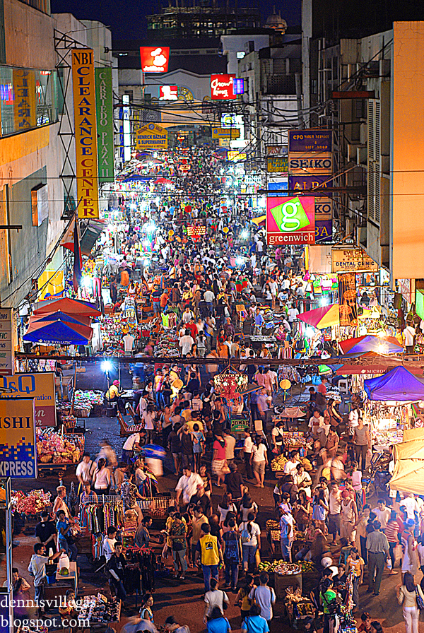
It's funny to think that with all the so-called "financial meltdowns" being experienced by many countries worldwide, the Philippines seemed to be relatively unaffected--judging by what you see in this picture. But this photo is misleading: not everyone here is shopping---many are just passers-by, hangers-on, pedestrians, and window shoppers.
For those who have money to spend this Christmas, many just buy things of superficial value and hardly necessary. Those who have credit cards tend to spend much more than they earn, and in the end suffer by paying endless mortgages. The higher the credit limit can actually boost more credit shopping, giving momentarily material satisfaction and living a life of worries and misery in the end. This is precisely the reason why I don't have credit cards. I have been approached several times by bank agents into getting one. Many lure me by offering freebies like watches, cellphones, etc., but I refused, since I only want to spend what is inside my pocket or bank.
As it happened, many credit card holders cannot pay back, and the banks, to protect their interests, declare bankruptcy. The government--fearing "financial meltdown" then helps by "bail-out" giving these banks financial help--literally billions from the taxpayers' money. In the end, it is us, the taxpayers, who have to pay the credits, while the bank executives still live in royal luxury with all their mansions and limousines.
Advance Merry and "credit-free" Christmas!
----------------------------------------------------------------
Poscript: You may have noticed that I am currently experimenting with htmls/xmls, skins, and templates of my blog. For the time-being, this look will be okay for now. All the sidebar links and archives are already placed at the bottom of this page. I guess I am not a fan of sidebars...so for the moment, they will be at the bottom, until such time that I can definitely decide which part of the page they should be placed. Again, sorry for this inconvenience!
Meningioma


Findings
CT shows a large circumscribed vertex mass in the right parietal region with heterogeneous hyperdensity and calcification. There is significant vasogenic edema and minimal if any mass effect or midline shift.
MRI from next day shows broad attachment of the lesion to the dura with moderate contrast enhancement. T2 prolongation compatible with vasogenic edema is again present.
Differential diagnosis:
- Meningioma
- Metastatic disease with hemorrhage and/or calcification
- GBM
- Low grade astrocytoma
- Angiosarcoma
- Tuberculoma
Diagnosis: Meningioma
Discussion
Meningiomas are thought to arise from arachnoid cap cells and may arise in the spinal cord or intracranially. Fewer than 10% are symptomatic. They may present with headache, seizure, or focal neurologic signs due to cranial nerve or brain parenchymal compression or vascular compression. Known causes include radiation and genetic abnormalities (including a relationship to NF2). Other causes are speculated as well. Meningiomas are generally considered benign tumors. However, a few histologic types can break this rule and invade cortex and even metastasize. Therapy includes conventional surgery and radiosurgery. Chemotherapy can be used following resection. Angiography is often performed for surgical planning and occasional embolization.
Radiologic overview of the diagnosis
Plain films of the skull may demonstrate hyperostosis and increased vascular markings. CT and MRI demonstrate extra axial, dural based lesions. Meningiomas typically enhance homogeneously and may have an enhancing dural tail (which may be more evident on coronal or sagittal MRI depending on the location)..On CT, the lesion may be isoattenuating to hyper attenuating but may contain calcifications. Vasogenic edema will likely be present and may be more apparent on MRI. T1 and T2 signal is variable. MR spectroscopy demonstrates a high alanine peak. Buckling of the cortex (seen in this case) is strongly suggestive of an extra axial lesion and should narrow the differential diagnosis. Other clues to extra axial location are brain cysts and trapped CSF. Angiographic findings include a sunburst vascular pattern and "mother-in-law" blush (comes early and stays late).

Monday, November 24, 2008
Sturge-Weber syndrome


Findings
Gyriform calcifications are observed over the left occipital lobe in the CT exam. The MRI demonstrates atrophy of the left cerebral hemisphere. There is enlargement of the left choroid plexus which demonstrates homogeneous enhancement post contrast. There is also gyriform enhancement post contrast most significantly on the left occipital lobe. There is diffuse enhancement of the subcutaneous tissues over the left eye.
Diagnosis: Sturge-Weber syndrome
Key points
Classically the patients have a facial port-wine stain, ipsilateral intracranial abnormalities, contralateral hemiparesis, hemiatrophy, mental retardation, and homonymous hemianopia. The severity of these features varies widely patient to patient. Commonly the patients will have glaucoma on the affected side. Seizures are also very common.
Only 8% of patients with port-wine stains have Sturge-Weber Syndrome. 13% of Sturge-Weber syndrome patients do not have a facial angioma.
There is no clear genetic link at this time. There is no sex or race predilection and it is very seldom seen more than once in the same family. Several different chromosomal abnormalities have been implicated.
Radiographically one can see "tram track calcifications" which are leptomeningeal calcifications like those seen on the CT image.
MRI can demonstrate the angiomatous abnormalities. In this case the cutaneous capillary angioma (port-wine stain) is well demonstrated as is the meningeal angiomatosis over the left occipital lobe. Cerebral hemiatrophy is well demonstrated by MRI as is the choroidal angiomatosis.
Multiple therapies are employed in these patients. The port-wine stains can be "removed" with laser treatments. Seizures can be treated with anticonvulsants. Refractory seizures can be treated surgically. The surgeries can be as extensive as a hemispherectomy.

Intracranial chondrosarcoma


Findings
Figure 1: Sagittal T1 demonstrates a low-signal mass centered in the sphenoid bone and extending into the anterior cranial fossa. The pituitary and suprasellar cistern are preserved.
Figure 2: Coronal T2 image shows displacement of the frontal lobes without brain edema confirming extra-axial location. The mass is high signal on T2 images which is non-specific but typical of chondrosarcoma. There is edema in and around both optic nerves.
Figure 3 and Figure 4: T1 weighted fat-suppressed post gadolinium axial and coronal images demonstrate avid enhancement of the solid mass.
Figure 5 and Figure 6: Axial and coronal CT demonstrate the calcified matrix within the mass taking characteristic “ring and arc” shapes. The anterior and superior walls of the sphenoid sinus are eroded confirming the aggressiveness of the tumor.
Differential Diagnosis:
- Chondrosarcoma
- Meningioma
- Chordoma
- Metastasis
- Lymphoma
Diagnosis: Intracranial chondrosarcoma
Intracranial chondrosarcoma is a slow-growing, locally invasive, rare malignant neoplasm of cartilaginous origin which accounts for 0.15% of all intracranial tumors and most commonly affects the skull base. Often when encountered, other anterior skull base malignancies, such as meningioma, metastasis, chordoma, rhabdomyosarcoma, and lymphoma may be difficult to distinguish. However, recognizing the range of appearances of intracranial chondrosarcomas on various imaging modalities along with clinical, gross, and histological studies may allow for improvement in the diagnosis, evaluation, and management of this malignancy.
Primary chondrosarcoma is divided into multiple variants, depending on the location and histological characteristics. These include conventional, clear cell, myxoid, mesenchymal, extraskeletal, and dedifferentiated. Skull base chondrosarcomas are most commonly of the conventional type and occupy areas that include petrosal bone, temporoccipital bone, clivus, sphenoethmoidal complex, and less commonly the frontal, parietal, and ethmoidal bones. Conventional chondrosarcomas can be further classified into histological subtypes of grade I, grade II, and grade III, with grade I demonstrating the least malignant and aggressive potential. The mesenchymal variant is the most malignant of all types and typically presents in a younger population. This type has a predilection for dural and cerebral extension.
Intracranial chondrosarcomas have been shown to be minimally sensitive to conventional radiation therapy, thus requiring radical excision of the tumor for effective management. Therefore, diagnostic imaging with MRI and CT is significant for neurosurgical assessment of tumor invasion and its anatomic orientation with any surrounding vascular, bony, and neural structures. Radiological imaging has enhanced diagnosis as CT is useful in finely delineating tumor invasion, bony invasion, and abnormal “ring and arc” or stippled calcification typical of chondrosarcomas. The tumor is normally isoattenuated or hyperattenuated with some degree of heterogeneous enhancement. MRI with gadolinium allows for evaluation of significant vessels and nerves such as the carotid arteries and optic nerves that lie in the preferred area of tumor growth. Furthermore, the tumor appears with decreased signal on T1 weighted images and increased signal on T2-weighted images. MRI enhancement will be mild to moderate, typically described with a “honey-combing” appearance due to islands of cartilage. In addition, it has been demonstrated that there is only mild edema surrounding chondrosarcomas in contrast to other skull base malignancies. MRI perfusion has also been documented to be effective in differentiating chondrosarcomas from other anterior skull base malignancies by assessing the cerebral blood volume and perfusion. Tumor vascularity is variable depending on the histological type; however, conventional and mesenchymal chondrosarcomas are commonly hypovascular, which differentiates it from the significantly vascular meningioma and metastatic lesion.
Although rare, intracranial chondrosarcomas should be considered in the differential diagnosis of any skull base malignancy that causes cranial nerve deficits. Clinically, patients commonly present with headaches, tinnitus, dizziness, decreased sense of hearing, visual symptoms such as diplopia and other oculomotor disorders depending on the location of the mass. The mean age of patients with intracranial chondrosarcomas has been reported to be 37 without any gender predilection. Although conventional radiation has limited indication, there has been considerable debate regarding optimal treatment with proton radiotherapy in combination with surgical excision. The difficulty in evaluating the outcomes of these various treatment strategies for skull base chondrosarcomas stems from the fact that this is a rare malignancy.
A Chinese Funeral
 The Chinese funeral procession is an spectacle of pomp and circumstance. At the head of the procession is the cart carrying some of the material things of the dead--as these will be burned during the burial. There are miniature paper houses and cars especially made to be burned in the funeral. Kim, or Chinese dead money, are spread out in the procession and burned in the burial.
The funeral parade is led by the live band playing a funeral march. It is followed by the hearse and the grieving family. One can actually hear the wails of the mourners, competing with the loud funeral music. The family, who are the chief mourners, must wear white colors, all the male members with a white band around their heads, while the women wearing white veils. The other relatives and close friends of the deceased must follow the order of procession, each wearing a sash pinned diagonally across their bodies.
Before the burial, all mourners must walk around the open coffin, each holding incense to offer to the dead. They plant the incense in the make-shift altar where the picture of their departed one is placed. Foods are also offered to the dead.
The photos below are being posted strictly for this blog only. I have been granted the permission of the family to post these pictures here, and not anywhere else. I will not accompany the pictures with captions and will instead let the pictures speak for themselves.
 The funeral cart that carries the material possessions of the dead. Ironically, the cart has a sign that reads "Mabuhay".


 The family members and friends pay final homage to the dead before internment.
Subscribe to:
Posts (Atom)