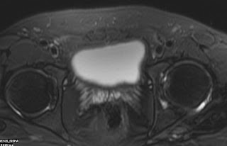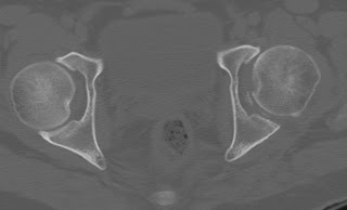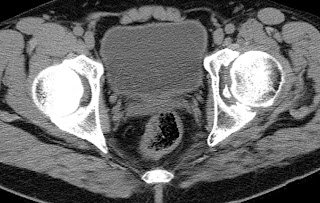


Here is a case of Acetabular fracture for the Radiology Grand Rounds submitted by Dr MGK Murthy, Dr Sumer Sethi of Teleradiology Providers. Concept and Archive of the Radiology Grand Rounds is available at- Radiology Grand Rounds.
This patient had posterior dislocation of hip reduced about 6 months back now he presents with complaint of pain in the left hip.
FAQ (questions to be answered)
(a)Is it unreduced?
NO
(b) Has it developed AVN?
NO
(c) Is there any associated injury which Xray did not pickup?
MRI is silent on it
(d) Can anything else help?
YES CT would help
(e) What does it show?
It shows post wall fracture comminuted with loose fragment in the joint cavity
(f)What are the complications of posterior dislocation hip?
Complications-- include avascular necrosis, osteoarthosis, sciatic nerve injury and heterotrophic ossification.
(g) What are the types of acetabular fractures?
Acetabular fractures are classified according to Judet classification usually.
Walls, Columns and Transverse varieties
Wall fractures
Anterior wall
Posterior wall
Posterior column with posterior wall (also a column fracture)
Transverse with posterior wall (also a transverse fracture)
Column fractures
Anterior column
Posterior column
Both-column
Posterior column with posterior wall (also a wall fracture)
Anterior column with posterior hemitransverse (also a transverse fracture)
Transverse fractures
Transverse
T-shaped
Transverse with posterior wall (also a wall fracture)
Anterior column with posterior hemitransverse (also a column fracture)
Common types (90%)
Both-column
Transverse with posterior wall
Posterior wall
T-shaped
Transverse
Hope you enjoyed this edition of Radiology Grand Rounds submissions are requested for the next Radiology Grand Rounds posted every month last sunday. If you interested in hosting any of the future issues contact me at sumerdoc-AT-yahoo-DOT-com.
No comments:
Post a Comment