
Welcome to the Second Edition of the monthly summary of the best in the Radiology Blogsosphere known as “ Radiology Grand Rounds”. Grand Rounds is an old tradition that doctors have. Once a week, they get together and talk about one case in detail. Keeping up with this tradition this Carnival of Medical Imaging has been named “Radiology Grand Rounds”. Every physician would agree that Subspecializtion is the need of the hour in medical field, hence the concept of a specialized Radiology Grand Rounds. Radiology Grand Rounds will be hosted on last Sunday of each month, the schedule and archive will be available at- Radiology Grand Rounds I would like to thank all the contributors for this edition of Radiology Grand Rounds.
WELCOME TO THE RADIOLOGY DEPARTMENT

THE RADIOGRAPHY & INVESTIGATIONS DEPARTMENT
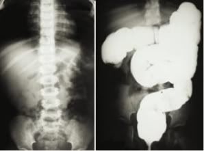
Jon Mikel of Unbounded Medicine presents a Case of Intussusception with Characteristic Radiological Pictures .
Intussusception is the invagination of a part of the intestine into itself, in other words is the prolapse of one part of the intestine into the lumen of an immediately adjoining part. It is the most common abdominal emergency in early childhood.
Radiological Findings- The abdominal plain film may be helpful because they may show frank intestinal obstruction or massively distended loops of bowel with absence of colonic gas. The ultrasound can be useful also, with a sensitivity and specificity approach 100%. The classic finding is a “bull’s eye” or “coiled spring” lesion (see below) representing layers of the intestine within the intestine.
DIGITAL RADIOGRAPHY DEPARTMENT

Kim of the Emergiblog writes on a Nurses' perspective on Computed or Digital Radiology. It goes on like-
Now this may be old hat to all you radiology-inclined folks, but we have a fantastic new system that has done away with the old x-ray film.X-rays are now viewed at a central station where the doctor sits via a computer screen.No more waiting for films to be developed and if the patient needs a copy of the x-ray, it can be placed on a CD that can be read on any computer.
COMPUTED TOMOGRAPHY DEPARTMENT
Bhavin Jhankaria has a post showing CT images of Emphysematous pyelonephritis The diagnosis on CT is relatively easy, with the presence of a focal area of necrosis and altered density with air, which may extend into the peri-renal space.
MRI DEPARTMENT
Bhavin Jhankaria has another submission from his Cardiac CT Blog pointing to an article by Timo Baks et al from Erasmus in Rotterdam, in the July issue of JACC, which shows that delayed enhanced, multislice CT (DE-MSCT) is as good as delayed hyperenhanced MRI (DE-MRI) in the assessment of infarction.
INTERESTING CASE DISCUSSION SECTION

Anil Aggarwal gives us a Book Review of Who Killed King Tut? Using Modern Forensics To Solve A 3,300-Year-Old Mystery by Michael R. King and Gregory M. Cooper, where Radiological methods are used to solve a famous Historical Mystery.
I would also like to point towards one of my famous previous post King Tut's CT scan rules out violent death which will act as a sequelae to this book. Recently a CT scan was done on King tut which revealed that there is no evidence of Head Injury!! So the mystery is finally solved or is it?
There is no doubt that the discovery of the sarcophagus of King Tutankhamen (rather irreverently referred to as "King Tut" in this book,.. or so it may seem) was one of the greatest historical discoveries of all time. Most readers would be familiar with the fascinating story of Howard Carter 's 1922 discovery of the tomb and the remains of the boy king whose gold funeral mask still continues to fascinate visitors to the Cairo Museum . Most, including this reviewer, had assumed that Tutankhamen died of natural disease. Several Egyptian mummies show the unmistakable scars of smallpox and tuberculosis. It therefore comes as a surprise to discover that he was murdered! Fascinated by the brief life and premature death of young King Tutankhamen, the authors of this book set off to unravel a so-called ancient mystery "using a combination of modern forensic archaeological evidence, modern forensic techniques, and psychological profiling" to determine whether or not young King Tutankhamen was actually murdered.
INTERVENTIONAL RADIOLOGY DEPARTMENT
The patient initially has a large left sided pleural effusion, then a smaller (by 3 litres)
left sided pleural effusion and a pneumothorax.
Filmjacket.com has a post showing images of
Filmjacket.com has a post showing images of
RF ablation of a hepatic cholangiocarcinoma metastasis. Cool images!!
That wraps up this month's highlights of the Radiology blogosphere. Hope the readers enjoyed the second edition of the Radiology Grand Rounds. If you liked any of these blogs, keep visiting them. Please email me at sumerdoc@yahoo.com if you are interested in hosting future Radiology Grand Rounds. Archive for the Radiology Grand Rounds here-Radiology Grand Rounds.
That wraps up this month's highlights of the Radiology blogosphere. Hope the readers enjoyed the second edition of the Radiology Grand Rounds. If you liked any of these blogs, keep visiting them. Please email me at sumerdoc@yahoo.com if you are interested in hosting future Radiology Grand Rounds. Archive for the Radiology Grand Rounds here-Radiology Grand Rounds.
For More updates on Radiology Grand Rounds A new discussion Group
has been created here, send me a mail to be invited to the group.
Group name: Radiology Grand Rounds
Group home page: http://groups.google.co.in/group/radgrandrounds
Group email address radgrandrounds@googlegroups.com
Be sure to tune in Next Month Last Sunday 27th August, when Grand Rounds
Be sure to tune in Next Month Last Sunday 27th August, when Grand Rounds
No comments:
Post a Comment