Saturday, July 29, 2006

Filipino Street Children


RADIOLOGY GRAND ROUNDS-II

Photobucket - Video and Image Hosting


Welcome to the Second Edition of the monthly summary of the best in the Radiology Blogsosphere known as “ Radiology Grand Rounds”. Grand Rounds is an old tradition that doctors have. Once a week, they get together and talk about one case in detail. Keeping up with this tradition this Carnival of Medical Imaging has been named “Radiology Grand Rounds”. Every physician would agree that Subspecializtion is the need of the hour in medical field, hence the concept of a specialized Radiology Grand Rounds. Radiology Grand Rounds will be hosted on last Sunday of each month, the schedule and archive will be available at- Radiology Grand Rounds I would like to thank all the contributors for this edition of Radiology Grand Rounds.


WELCOME TO THE RADIOLOGY DEPARTMENT


Photobucket - Video and Image Hosting


THE RADIOGRAPHY & INVESTIGATIONS DEPARTMENT
Photobucket - Video and Image Hosting


Jon Mikel of Unbounded Medicine presents a Case of Intussusception with Characteristic Radiological Pictures .
Intussusception is the invagination of a part of the intestine into itself, in other words is the prolapse of one part of the intestine into the lumen of an immediately adjoining part. It is the most common abdominal emergency in early childhood.
Radiological Findings- The abdominal plain film may be helpful because they may show frank intestinal obstruction or massively distended loops of bowel with absence of colonic gas. The ultrasound can be useful also, with a sensitivity and specificity approach 100%. The classic finding is a “bull’s eye” or “coiled spring” lesion (see below) representing layers of the intestine within the intestine.


DIGITAL RADIOGRAPHY DEPARTMENT

Photobucket - Video and Image Hosting


Kim of the Emergiblog writes on a Nurses' perspective on Computed or Digital Radiology. It goes on like-
Now this may be old hat to all you radiology-inclined folks, but we have a fantastic new system that has done away with the old x-ray film.X-rays are now viewed at a central station where the doctor sits via a computer screen.No more waiting for films to be developed and if the patient needs a copy of the x-ray, it can be placed on a CD that can be read on any computer.


COMPUTED TOMOGRAPHY DEPARTMENT


Bhavin Jhankaria has a post showing CT images of Emphysematous pyelonephritis The diagnosis on CT is relatively easy, with the presence of a focal area of necrosis and altered density with air, which may extend into the peri-renal space.


MRI DEPARTMENT

Photobucket - Video and Image Hosting


Bhavin Jhankaria has another submission from his Cardiac CT Blog pointing to an article by Timo Baks et al from Erasmus in Rotterdam, in the July issue of JACC, which shows that delayed enhanced, multislice CT (DE-MSCT) is as good as delayed hyperenhanced MRI (DE-MRI) in the assessment of infarction.


Now MRI can be used in Utero itself!!
Sumer’s Radiology Site points to an Article

INTERESTING CASE DISCUSSION SECTION

Photobucket - Video and Image Hosting


Anil Aggarwal gives us a Book Review of Who Killed King Tut? Using Modern Forensics To Solve A 3,300-Year-Old Mystery by Michael R. King and Gregory M. Cooper, where Radiological methods are used to solve a famous Historical Mystery.
I would also like to point towards one of my famous previous post
King Tut's CT scan rules out violent death which will act as a sequelae to this book. Recently a CT scan was done on King tut which revealed that there is no evidence of Head Injury!! So the mystery is finally solved or is it?
There is no doubt that the discovery of the sarcophagus of King Tutankhamen (rather irreverently referred to as "King Tut" in this book,.. or so it may seem) was one of the greatest historical discoveries of all time. Most readers would be familiar with the fascinating story of Howard Carter 's 1922 discovery of the tomb and the remains of the boy king whose gold funeral mask still continues to fascinate visitors to the Cairo Museum . Most, including this reviewer, had assumed that Tutankhamen died of natural disease. Several Egyptian mummies show the unmistakable scars of smallpox and tuberculosis. It therefore comes as a surprise to discover that he was murdered! Fascinated by the brief life and premature death of young King Tutankhamen, the authors of this book set off to unravel a so-called ancient mystery "using a combination of modern forensic archaeological evidence, modern forensic techniques, and psychological profiling" to determine whether or not young King Tutankhamen was actually murdered.

INTERVENTIONAL RADIOLOGY DEPARTMENT

Barbados Butterfly’s submission is a post with a
The patient initially has a large left sided pleural effusion, then a smaller (by 3 litres)
left sided pleural effusion and a pneumothorax.


Filmjacket.com has a post showing images of
RF ablation of a hepatic cholangiocarcinoma metastasis. Cool images!!


That wraps up this month's highlights of the Radiology blogosphere. Hope the readers
enjoyed the second edition of the Radiology Grand Rounds. If you liked any of these blogs, keep visiting them. Please email me at sumerdoc@yahoo.com if you are interested in hosting future Radiology Grand Rounds. Archive for the Radiology Grand Rounds here-Radiology Grand Rounds.


For More updates on Radiology Grand Rounds A new discussion Group
has been created here, send me a mail to be invited to the group.

Group name: Radiology Grand Rounds
Group email address radgrandrounds@googlegroups.com

Be sure to tune in Next Month Last Sunday 27th August, when Grand Rounds
will be hosted by Me again at Sumer's Radiology Site

Wednesday, July 26, 2006

Ganglioglioma


Findings

Within the medial subcortical portion of the anterior right temporal lobe, there is a 1.5 x 1.6 x 1.3 cm intraaxial lesion that demonstrates increased signal intensity on T2 and FLAIR images (Figure 2, 3, 5 and 6) and relatively low-signal intensity on T1-weighted images (Figure 1). The lesion has well-circumscribed and slightly-lobulated margins. No significant perilesional edema.
Postcontrast images do not reveal any appreciable enhancement (Figure 4 and 7).
This lesion appears to be located inferior and mostly anterior to the right amygdala, and expands the right parahippocampal gyrus medially and the right fusiform gyrus inferiorly.


Diagnosis: Ganglioglioma


Ganglioglioma is a well-differentiated, slowly growing neuroepithelial tumor composed of neoplastic neuronal and glial elements. Majority of gangliogliomas (85%) are low grade (WHO I). It occurs most commonly in children and young adults. Patients usually have a long-standing history of seizures and headaches.

Gangliogliomas are most commonly located in the superficial cerebral hemispheres, especially the temporal lobes. These tumors are usually firm, well-circumscribed masses and may expand the involved cortex. On CT, these have an appearance of low-density or cystic masses. Focal enhancement is seen in 50% of the cases and calcification in approximately 35%. MRI demonstrates these hemispheric lesions in a cortical location, being hyperintense on T2- and hypointense on T1-weighted images. Gadolinium enhancement is variable, often focal or nodular. PET typically shows decreased activity, indicating tumor hypometabolism. In children under 10 years, gangliogliomas can be larger and more cystic.

These tumors have an excellent prognosis with complete surgical excision. The majority (80%) of the patients are seizure-free after surgery. Malignant transformation of ganglioglioma is unusual.

Should we use MRI for Fetal Spine Imaging?


"In-utero MR imaging (iuMR) has entered the clinical arena during the last decade. It is used mainly for imaging fetal brain abnormalities. Authors report their experience of imaging the fetal spine and spinal cord in fetuses with known or suspected abnormalities diagnosed on US imaging. In 40 (80%) of 50 fetuses, iuMR and US imaging were in complete agreement. In the other 10 fetuses (20%), iuMR provided additional information or changed the diagnosis, including 8 fetuses where the iuMR could find no abnormality and was found to be correct by later follow-up.
Authors conclude-The clinical impact of iuMR may be numerically less than with brain abnormalities, but is still sufficient to warrant its use, especially if there is any uncertainty about the US imaging, and particularly as a relatively high proportion of diagnoses on US imaging are false-positives."

Tuesday, July 25, 2006

Imaging Of Aortic Dissection

Reference-Diagnostic Accuracy of Transesophageal Echocardiography, Helical Computed Tomography, and Magnetic Resonance Imaging for Suspected Thoracic Aortic Dissection: Systematic Review and Meta-analysis. By Shiga T et al in Arch Intern Med 2006 Jul 10;166(13):1350-6.

"Patients with suspected thoracic aortic dissection require early and accurate diagnosis. Aortography has been replaced by less invasive imaging techniques including transesophageal echocardiography (TEE), helical computed tomography (CT), and magnetic resonance imaging (MRI); however, accuracies have varied from trial to trial, and which imaging technique should be applied to which risk population remains unclear.
In their study authors found Pooled sensitivity and specificity were comparable between imaging techniques. The pooled positive likelihood ratio appeared to be higher for MRI than for TEE or helical CT. All 3 imaging techniques, ie, TEE, helical CT, and MRI, yield clinically equally reliable diagnostic values for confirming or ruling out thoracic aortic dissection."

Sunday, July 23, 2006

My Recent Walk in Avenida


This is Avenida, one of the busiest boulevards in Manila. Here you will find all kinds of people, bartering, selling, or buying, or just strolling around looking for someone to love :P. I always walk along Avenida after my work, and not a few times was I approached by a girl or an older lady offering their bodies for a few hundred pesos. If you have a keen eye you can spot some of them in this picture.

This is Plaza Lacson in Sta. Cruz. This photo was taken from the LRT Carriedo Station in Sta. Cruz. The Old Prudential Bank Building is in the background. In front of it is the perrenial statue of Mayor Arsenio Lacson.

Friday, July 21, 2006

Submissions required for the Radiology GR-II


Dear Friends as you all know Second edition of the monthly summary of the Radiology Blogosphere is coming up on the last Sunday of this month, 30th July. So Hurry Up!! rush in your Radiology Related posts to me at-
sumerdoc-AT-yahoo-DOT-com
Please Note you need not be a Radiologist to post, I am looking forward to posts from all technicians, students, Physicians, nurse etc...
Details about future schedule and guidelines-
I'm back from a short trip from Samar. The plane left Calbayog at 7:00 in the morning and I arrived exactly 9:00 A.M. in Manila. I had wanted to see Mayon Volcano from the plane's window but the thick clouds hid not only Mayon, but also the rest of the more viewable things under our plane's wings.
I miss Manila so I decided to eat lunch in Ma Mon Luk. After the plane ride, I really don't want to line up and serve myself in fast foods. Ma Mon Luk has waiters and I can sit comfortably while seated on a chair. Nothing compares to the excitement of waiting for your order to arrive, and when it is finally served, nothing beats the aroma of steaming broth!

This is the regular menu in Ma Mon Luk. The food may be a little more expensive than McDonalds or Jolibee, but I tell you I will never trade Ma Mon Luk Siopao with the Big Mac. I really like that byline "Ang Mami dito ay unang imbento, Una sa Lasa, Una sa Sarap". Now that is really cool, doesn't it?

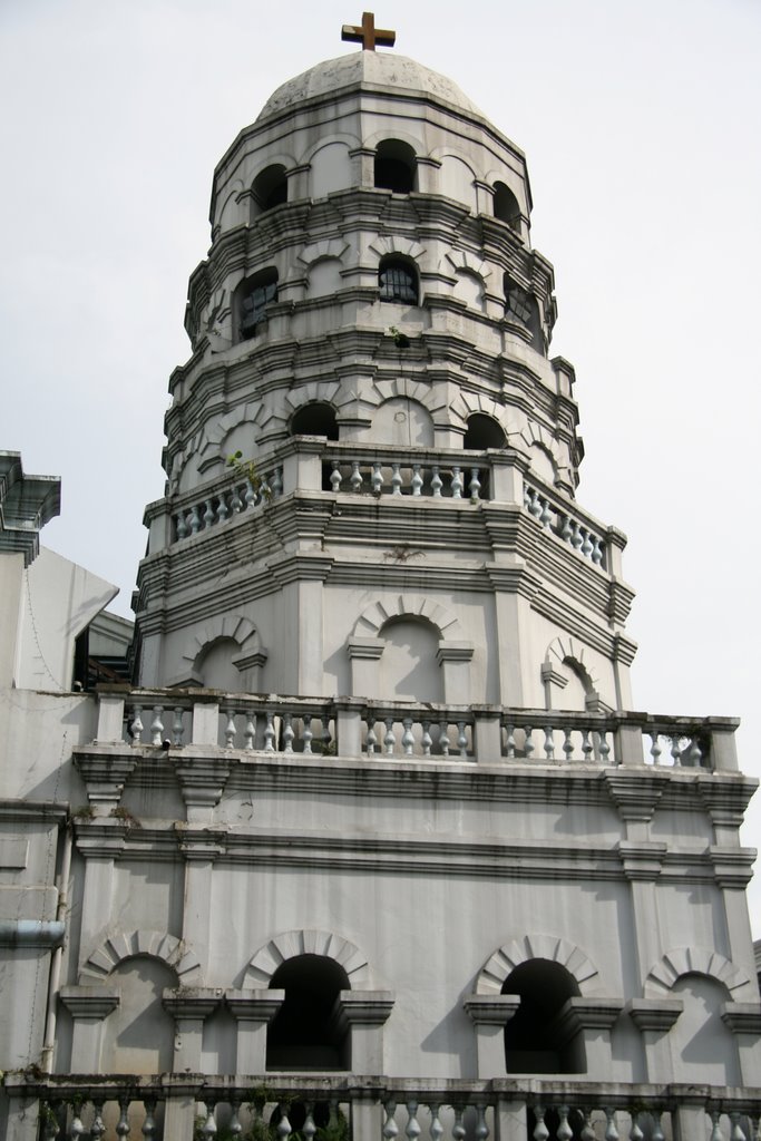
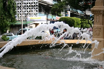
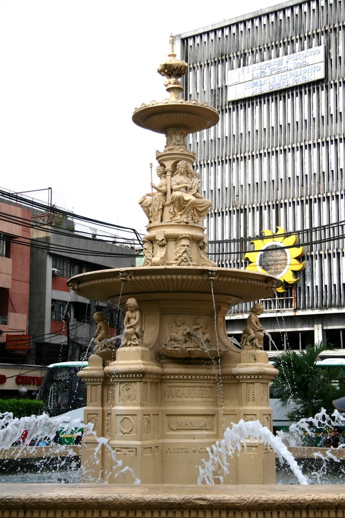
Ever since I had my Rebel XT, I just miss my little compact Sony DSC-T5. So I took it out of my pocket and exercised its legs.
It still takes good pictures despite its being 2 years old(an octogenarian for a camera).
And here are some of them:


Well, these guys are the waiters of Ma Mon Luk. All of them are regulars there, and some have been waitering there even when Ma Mon Luk was still living. They may be snobbish the first you meet them, but I tell you if you eat there, kindly leave 10 or 20 pesos in tip and the next time you eat there, they'll treat you like a king!

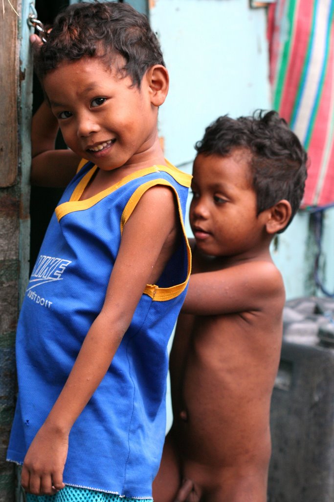
I'll post more pictures soon. Meanwhile it's time to take some rest...in the restroom.

Wednesday, July 19, 2006

Blogger Blocked In India-Violation of freedom to speak!!

I was not able to access my blog for last few days thought it was some problem with blogger, till i saw the following news item... Highly objectionable act by the Goverment.. straight violation of the right to freedom of speech...
Indian Government Blocks Web Access
Action Mirrors That of China (Source-Washington Post)
"India's Internet regulators have started blocking several Web sites, following the lead of China, where government censors heavily restrict the flow of online information. India's department of telecommunications sent an order late last week to Internet service providers to block several Web sites, according to a department spokesman. The spokesman, Rajesh Malhotra, declined to disclose the contents of the letter or discuss the order, saying it was a "confidential exchange of information between the department and the operators." Several telecom operators confirmed that they were directed to block more than 15 Web sites. Close to a third of those are home to blogs, or personalized Web logs, such as Blogger.com and Geocities.com."

Atypical meningioma


Findings

CT without contrast demonstrates an extra-axial heterogeneous mass with solid and cystic components in the left frontal lobe adjacent to the falx. There is calcification along its medial attachment with the falx.
Figure 2: CT with contrast demonstrates homogeneous enhancement of the solid component of the left frontal lobe mass.
Figure 3: Axial T1-weighted MRI with gadolinium contrast demonstrates homogeneous enhancement of the solid peripheral component.
Figure 4: Sagittal T1-weighted MRI demonstrates a hypointense mass in the left frontal lobe. There is curvilinear increased signal intensity along the periphery of the mass, which represents calcifications as demonstrated on CT.


Diagnosis: Atypical meningioma


World Health Organization (WHO) Classification of Meningiomas
- Meningioma (typical benign)
- Atypical meningioma
- Anaplastic (malignant) meningioma

The peak incidence of atypical meningioma occurs in the fourth decade, whereas benign meningiomas show a peak incidence in the fifth decade. Men are affected equally as women with atypical meningioma. In contradistinction, women are affected more frequently with benign meningioma. The most common anatomical location of atypical meningioma is in the parasagittal region (43.7%), followed by the cerebral convexities (15.6%). The recurrence rate of atypical meningioma is 28% within two years, compared to 9.3% for benign meningioma. Atypical meningiomas represent an intermediate category of tumor that has a higher relative risk for recurrence.


Neuroradiologic features of meningiomas

The typical meningioma is a homogeneous, hemispheric, markedly enhancing extraaxial mass located over the cerebral convexity, in the parasagittal region, or arising from the sphenoid wing. However, typical meningiomas and several histologic variants of meningioma can have unusual or misleading radiologic features that may not be suggestive of meningioma. Unusual imaging features, such as large meningeal cysts, ring enhancement, and various metaplastic changes (including fatty transformation), can be particularly misleading. Because meningiomas are so common, the radiologist must be aware of their less frequent and uncharacteristic imaging features in order to suggest the correct diagnosis in atypical cases.

Saturday, July 15, 2006

Cerebral Malaria-MRI

MR of cerebral malaria. In AJNR Am J Neuroradiol 1998 May;19(5):871-4. By Cordoliani YS et al.
"In three cases of cerebral malaria, MR imaging disclosed either cortical infarcts (one case) or hyperintense areas of white matter (two cases) on T2-weighted and fluid-attenuated inversion-recovery sequences. These white matter abnormalities were, in one case, sharply limited, symmetrical, hyperintense, and unenhanced; in the other case, they were diffuse, hyperintense, and had a more limited focus. The diffuse hyperintensity was probably due to edema, whereas focal lesions were probably associated with gliosis."

Renal Artery Stenosis-Anatomical Distribution

"Focal Renal Artery stenosis in their study group was distributed as follows 25% main renal artery, 50% 2nd order branch, 12.5% 3rd order branch, and 12.5% accessory renal artery. They concluded Hypertensive children without comorbid conditions who have RAS usually have single, focal branch artery stenoses. This distribution supports angiography in these patients because of its superior sensitivity in detecting branch vessel disease and its therapeutic role in percutaneous transluminal renal angioplasty."

Tuesday, July 11, 2006

Some useful Radiology RSS Feeds

Found some useful Radiology Related RSS feed which Radiologists can add to their RSS readers and keep updated to the latest in Radiology.

Sumer's Radiology Site
http://sumerdoc.blogspot.com/atom.xml

Journal of Computer Assisted Tomography
http://www.jcat.org/pt/re/jcat/toccurrentrss.xml


Topics in Magnetic Resonance Imaging
http://www.topicsinmri.com/pt/re/tmri/toccurrentrss.xml


Journal of Thoracic Imaging
http://www.thoracicimaging.com/pt/re/jti/toccurrentrss.xml


Medscape Radiology Headlines
http://www.medscape.com/cx/rssfeeds/radiology.xml

Friday, July 7, 2006

Availability of Sonography in Emergency Gynecology Unit useful

In an Article entitiled "Impact of the availability of sonography in the acute gynecology unit." in Ultrasound Obstet Gynecol 2006 Jun 29; [Epub ahead of print] by Haider Z et al, the authors investigated the impact of the availability of transvaginal sonography at the time of initial assessment of the emergency gynecology patient. The initial assessment of acute gynecology patients is usually based on history and clinical examination and does not involve ultrasound. Authors found that Following the ultrasound examination there was a change in clinical management for 38.1% of non-pregnant women and a reduction in admissions (from 37.1% to 19.4%) and outpatient follow-up examinations (from 25.7% to 18.1%). It appears that the availability of transvaginal sonography at the time of initial assessment of emergency gynecology patients improves diagnostic accuracy and reduces unnecessary admissions and follow-up examinations.

Interesting post on Desert Imaging Blog

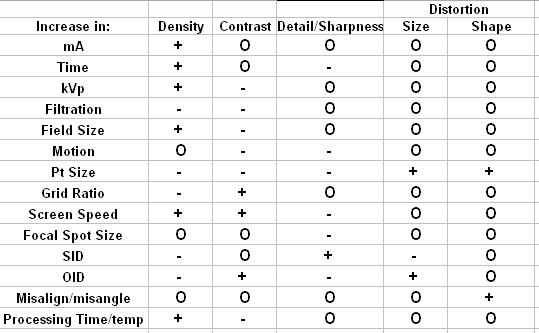
+ = increase, - = decrease, O = no change

Interesting post in Desert Imaging in which he talks about a Real Tough Radiography examination he appeared.
He goes like-
"It was one of those tests with questions like:"If you increase focal spot size how does it affect image detail?", "If kVP is increased by 15% what will happen to image density?", "When you increase OID what happens to radiographic contrast?" yada, yada, yada, blah, blah, blah, etc., etc., etc......."
And then follows up with a very useful Table for all Radiographers as well as Radiology Residents...

Thursday, July 6, 2006

Overdiagnosis Of Breast Cancer

Overdiagnosis and Overtreatment of Breast Cancer: Is Overdiagnosis an Issue for Radiologists?
Complete article here at Medscape
"Overdiagnosis is diagnosis of cancers that would not present within the life of the patient and is one of the downsides of screening. This applies to low-grade ductal carcinoma in situ and some small grade 1 invasive cancers. Radiologists are responsible for cancer diagnosis, but at the time of diagnosis they cannot determine whether a particular low-grade diagnosis is one to which the definition of overdiagnosis applies. Overdiagnosis is likely to be driven by technological developments, including digital mammography, computer-aided detection and improved biopsy techniques. It is also driven by the patient's fear that cancer will be missed and the doctor's fear of litigation. It is therefore an issue of importance for radiologists, presenting them with difficult fine-tuned decisions in every assessment clinic that are ultimately counted later by those who evaluate their screening."

Monday, July 3, 2006

Future of Double Contrast Barium Enema in Question

In a Study by Ferrucci JT, Double-contrast barium enema: use in practice and implications for CT colonography in AJR Am J Roentgenol 2006 Jul;187(1):170-3

"This study examines the use and yield of double-contrast barium enemas (DCBEs) for colorectal polyp detection in current clinical practice outside the research setting. A total of 244 out of 665 (36.7%) barium enema studies were performed using the double-contrast technique over the 4-year period-that is, approximately one per week. The most common indication for a DCBE (109/244 or 44.6%) was to complete a failed, incomplete, or inconclusive colonoscopy.Overall, only 14 of the 244 (5.7%) studies gave positive reports for polyps, and of these, five were shown to be false-positive at later colonoscopy. Only six polyps 10 mm or larger were positively detected during the entire study, which is approximately one per 60 studies or one every 8 months
Authors concluded that in their centre a DCBE is a low-yield procedure for detecting polyps, with a high false-positive rate, and is not likely to be performed by experienced practitioners in the future."