"The aim of this website is to be a completely free radiology education resource. Dr Simon has developed software which allows the user to view entire CT and MRI datasets over a broadband connection - in order to be able to test themselves in a real life environment - rather than with static images forun in many teaching websites. "
Wednesday, January 31, 2007
Off for Now!
Thank you for visiting my blog. I am taking a temporary leave from the blogging world because I will take time off to spend vacation in the southern provinces.
Also my computer got virused and all my recent precious photos of Cubao and Manila got erased from my hard drive. I did not keep a back up. My Calbayog photos survived though, and I'm happy just for that.
When I come back, I'll bring you more pictures of the places I've been to.
Bye for now!
Also my computer got virused and all my recent precious photos of Cubao and Manila got erased from my hard drive. I did not keep a back up. My Calbayog photos survived though, and I'm happy just for that.
When I come back, I'll bring you more pictures of the places I've been to.
Bye for now!
Arachnoid cyst


Findings
There is a small left frontal extra-axial hypodense fluid collection with associated thinning of the calvarium. No significant mass effect on the brain parenchyma. No acute intracranial abnormalities.
Differential diagnosis:
- Arachnoid cyst
- Chronic subdural hematoma
- Subdural hygroma
- Epidermoid cyst
- Prominent CSF space
- Porencephalic cyst
- Neuroglial cyst
Diagnosis: Arachnoid cyst
Key points
An arachnoid cyst is a benign, intra-arachnoid, CSF-filled cyst that does not communicate with the ventricular system. Arachnoid cysts are fairly common, accounting for approximately 1% of all intracranial masses. They are often asymptomatic and are found incidentally, but are twice as common in imaging performed for history of seizure. If present, symptoms vary with the size and location of the cyst and can include, headache, sensorineural hearing loss, dizziness, and even obstructive hydrocephalus. Arachnoids cysts are usually stable in size but have been known to slowly enlarge. No treatment is usually required, but resection, fenestration, or shunt placement may be performed for serious symptoms.
Radiology
In general: Arachnoid cysts may appear anywhere in the cranium, although the middle cranial fossa is the most common location. They can vary in size from only a few millimeters to ten centimeters or more. They typically displace cortex and may even "buckle" the gray-white border.
CT: CSF density, usually associated thinning/remodeling of adjacent bone, no enhancement with contrast
MR:
- T1WI - isointense with CSF, sharp margins
- T2WI – isointense with CSF
- FLAIR – suppresses completely
- DWI – no restricted diffusion
- T1+C – no enhancement
Monday, January 29, 2007
Lemen Law 2007
Ok, I'll give this blog thing another shot. I might move it soon, seeing that MySpace is trying to make money off of the people who write blogs on their site by putting Google Adwords ads below the blogs. Power to the people! Down with the man!
Colleen and I were talking a little bit about religion the other night, and started talking about those hardcore religious zealots that re-elected George W. Bush, and how those hardcore religious zealots are primarily in the middle states and have not seriously infected the liberal east and west coasts....
Well, they're heeeeeeeeeeerrrre.......
I'm all for free speech, but I love stories about crazy old women more. Especially if the crazy old women live about ten miles from me.....
Anne Lemen, a Balboa Island resident, is bringing her case for free speech to the California Supreme Court. An innocent Christian woman, just trying to make the world a better place, right?. She wants the world to know that the Village Inn on Balboa Island, is mafia, as are her former husband and the guards at a local church, and the police are in cahoots. Oh yeah, and she claims that the Village Inn has tried to murder her.
Ms. Lemen has reportedly videotaped the Village Inn's customers, told patrons that the bar makes sex videos, dabbles in child pornography, distributes illegal drugs, encourages lesbian activities, has mafia links, is a whorehouse and sells tainted food.
By the way, Colleen, make reservations at the Village Inn on Balboa Island for about 8:00 tonight.....
The owner has said that she once parked in front of his restaurant and blasted the horn for 30 minutes nonstop, and on another evening he watched as passersby scanned his window menu and fled after Lemen told them the food was poisonous and the place was filled with rats.
Lemen reportedly told her interviewer, "There have been four attempts on my life, and the police refuse to investigate because they are part of the bar."
Well, a judge has ordered her to stop bugging the patrons and making accusations that the Village Inn is not a reputable establishment.
Her recent appeal is taking her case for free speech to the Supreme Court of California. Her Christian friends describe her as a cross between Florence Nightingale, Joan of Arc and Erin Brockovich.
To add to Ms. Lemen's crazy religious zealot status:
1. Her garden contains a large stone Bible with Christian verses and a small lighthouse with the words "The Lord Is Our Light." Little booklets about Christianity, titled "Smile, Jesus Loves You," are there for passersby to pick up.
2. She also hangs a large silver circle emblazoned with an eagle and the words "In God We Trust." She said she put it up after hearing that civil libertarians opposed the mention of God on money.
3. Lemen popped a videotape into her VCR for the reporter, to show scenes of crowds of young adults in the early-morning hours outside the bar several years ago. A young woman in the crowd wore a halter top.
"Look at how she dresses," Lemen gasped.

4. She puts on her crown and holds her torch high, as she puts her arm around her front yard Statue of Liberty (holding a Holy Bible) and belts out religious hymns for passersby.
Well, it is interesting to note that Ms. Lemen is waiting for her appeal in a gated, adult community miles from Balboa Island.
I'll try to keep everbody updated on "Lemen Law 2007"!
Lumbar Disc Pathology-Classification
Here is an excellent link for those wanting to learn how to report MRI in degenerative spine by
Dr Peng Hui LEE
Clinical Director of Radiology, Mid Essex Hospitals NHS Trust
Nomenclature and Classification of Lumbar Disc Pathology
Dr Peng Hui LEE
Clinical Director of Radiology, Mid Essex Hospitals NHS Trust
Nomenclature and Classification of Lumbar Disc Pathology
Vein of Galen aneurysm


Findings
There is a large vascular structure just posterior to the third ventricle and superior to the cerebellar vermis. There is some mass-effect on the third ventricle with dilatation of the third ventricle and frontal horns. No definite intracranial hemorrhage noted. MRI shows a large tubular structure with flow identified in the region of the vein of Galen extending into the straight sinus towards the torcula Herophili. There are numerous collateral vessels within the ambient and perimesencephalic cisterns as well as surrounding the brainstem.
Diagnosis: Vein of Galen aneurysm
Key points
Vein of Galen "aneurysm" is actually an AVM with an associated varix of the vein of Galen.
Two main types exist
- Vein of Galen AVM.
- Vein of Galen aneurysmal dilatation.
Vein of Galen AVM is the result of a developmental malformation that involves the vein of Galen. It is an A-V shunt in the wall of an embryologic venous precursor (Median Vein of the Prosencephalon [MVP]) and in this situation a normal vein of Galen does not exist and is replaced by a persistent MVP. The MVP does not drain normal brain tissue. In other words, a Vein of Galen AVM is not anatomically associated with a parenchymal AVM. The dilated MVP drains via the dural sinuses. Vein of Galen AVM presents in infancy with high output cardiac failure and macrocephaly. The A-V fistulas may be single or multiple. Intrauterine diagnosis is usually possible by doppler. Intracranial hemorrhage is rare.
Vein of Galen AVM should be differentiated from aneurysmal dilatation of the vein of Galen which occurs in association with a parenchymal AVM or the rare asymptomatic varicose vein of Galen.
Therapy: Endovascular embolization via an arterial or venous access.
Radiology
Transcranial doppler sonogram shows an anechoic structure in the superoposterior aspect of the third ventricle with flow.
CT, MRI and MRA show the vascular abnormality and assess the brain parenchyma for abnormalities.
Conventional angiography is essential to assess the malformation and to plan the therapy.
Sunday, January 28, 2007
Radiology Grand Rounds-VIII
Welcome to the VIII Edition of the monthly summary of the best in the Radiology Blogsosphere known as “ Radiology Grand Rounds”. Grand Rounds is an old tradition that doctors have. Once a week, they get together and talk about one case in detail. Keeping up with this tradition this Carnival of Medical Imaging has been named “Radiology Grand Rounds”. Every physician would agree that Subspecializtion is the need of the hour in medical field, hence the concept of a specialized Radiology Grand Rounds. Radiology Grand Rounds will be hosted on last Sunday of each month, the schedule and archive will be available at- Radiology Grand Rounds. I would like to thank all the contributors for this edition of Radiology Grand Rounds.
This Edition we will start by informing my readers that we are getting famous, yes the concept of Grand Rounds including Radiology Grand Rounds was featured in Indian Pediatrics current edtion. Indian Pediatrics is one of the top most Indian Indexed Journal.
Anybody who knows about Radiology would know that one of the key arts in the field of diagnostic imaging is writting a proper report which conveys the impression to the referring clinician, i think everyone would agree writting a good report is very crucial to a successful radiology practise. Filmjacket.com has sent a link to a new database of radiology report templates. It was just placed online and will soon be populated with more report templates. It already has few templates on Ultrasound, rest of the categories are coming up. Good effort.
Filmjacket.com also presents an image gallery of various presentations of tuberculosis. Amazing collection of skeletal tuberculosis at various sites. Title of the series-TB or not TB submitted to them by Dr Priya Chudgar.
Odysseys of George submits to us a very interesting case of a 72 year old lady who presented with anaemia. Look at the images findings are obvious and discussions following the images are very interesting. My Tip-try making the diagnosis before reading the entire discussion.
And another case from him aptly titled breathing on bended tube, this is a case of an elderly man referred for loss of weight, stridor(noisy, harsh breathing) for the last one year which worsened over the last 2 months.
Dr MGK Murthy (Sr Consultant) at Teleradiology Providers talks about differential diagnosis of a supraclinoid aneurysm. He says at times "Meningimas which are cystic, haemangioblstomas and pilocytic astrocytomas could present a diagnostic difficulty.However intense nodular enhancement, lack of perilesional oedema, chronic headache of long duration with no deficit are supportive of aneurysm"
Hot Discussion Topics From Sumer's Radiology Site
(comments and suggestions on each topic are welcome)
That wraps up this month's highlights of the Radiology blogosphere. Hope the readers enjoyed the VIII edition of the Radiology Grand Rounds. If you liked any of these blogs, keep visiting them. Please email me at sumerdoc@yahoo.com if you are interested in hosting future Radiology Grand Rounds. Also visit our sister concern Teleradiology Providers. Archive for the Radiology Grand Rounds here-Radiology Grand Rounds. Be sure to tune in Next Month Last Sunday 25th Feb, when Grand Rounds will be hosted at- Cochin Blogs mail to-drjoea@gmail.com or sumerdoc@yahoo.com
Thursday, January 25, 2007
Radiology Grand Rounds feature in Indian Pediatrics
Indian Pediatrics Journal in its Pedscapes Column by Dr Sidharth, a fellow blogger from India who runs Pediatricsinfo.com features the concept of Grand Rounds. Here is the link to the full article. It talks about Medical, Pediatrics, Radiology and Nursing Grand Rounds.
Reference- Concept of Online Grand Rounds (Pedscapses). Indian Pediatrics 2007; 44:58.
Nevoid Basal Cell Carcinoma (Gorlin) Syndrome (NBCCS)


Findings
Figure 1: Characteristic calcification of the petroclinoid ligaments and the diaphragma sellae is present.
Figure 2: In addition, there are dural calcifications involving the falx cerebri and tentorium cerebelli.
Figure 3: Soft tissue lesion involving the right scalp is also visualized and corresponds to pathologically proven basal cell carcinoma.
Figure 4: There is subtle parallel orientation of the bodies of the lateral ventricles that suggests dysgenesis of the corpus callosum, a finding that may occur in as many as 10% of patients with NBCCS.
Diagnosis: Nevoid Basal Cell Carcinoma (Gorlin) Syndrome (NBCCS)
NBCCS (Gorlin syndrome) is a rare autosomal dominant disorder characterized by multiple basal cell carcinomas, odontogenic keratocysts of the jaw, palmar/plantar pits, calcification of the falx cerebri, and rib and spine anomalies. Patients with NBCCS have a propensity to develop multiple neoplasms such as basal cell carcinoma (>90%), medulloblastoma (10%), meningioma, and ovarian or cardiac fibromas.
Radiologic evaluation is important in patients suspected of having NBCCS because the identification of a pathogenic mutation is not always possible, and the clinical manifestations may be subtle, especially in children or African Americans. Recognition is also clinically important as these patients are sensitive to ionizing radiation and have been known to develop iatrogenic malignancy as a result of radiation therapy.
Key radiologic features include:
- Extensive calcification of the falx cerebri, tentorium cerebellum, diaphragm sella, or petroclinoid ligaments
- Odontogenic keratocyst of the jaw
- Bifid, splayed, fused, or absent ribs, especially involving the 3rd to 5th ribs
- Scoliosis, hemivertebrae, or fused vertebrae
- Flame-shaped lucencies of the hands or feet
- Scapular abnormalities
Nevoid Basal Cell Carcinoma Syndrome includes two Major critera or one Major and two Minor criteria
Major Criteria
- More than two basal cell carcinomas or one basal cell carcinoma under the age of 20
- Odontogenic keratocysts of the jaw
- Three or more palmar or plantar pits
- Lamellar calcification of the falx cerebri
- Rib Anomalies (bifid, synostosed, hypoplastic)
- First degree relative with NBCCS
Minor Criteria
- Spina bifida occulta or other vertebral anomalies
- Brachymetacarpaly in at least one limb
- Hypertelorism or telcanthus
- Frontal bossing
- Sprengel deformity
- Ovarian fibroma
- Medulloblastoma
- Meningioma
- Flame shaped lucencies of the phalanges
- Bridging of the sella turcica
- Mesenteric cysts
- Cardiac fibroma
- Dysgenesis/agenesis of the corpus callosum
Wednesday, January 24, 2007
AJR & Rejection of Manuscripts
"Countries in which English is the primary language had higher acceptance rates than those in which English is not the primary language. Countries with English as the primary language, including Canada, the United Kingdom, and Australia, had rejection patterns similar to that of the United States. Language problems were not a major reason for rejection, except for manuscripts from China. Lack of new or useful knowledge was by far the most common reason for rejection in all countries. High-quality scientific work is key to overcoming barriers to publication. Designing an appropriate study that answers a clearly defined and pertinent question is an important first step. Language problems were not a major cause of rejection, except for manuscripts from China. "
Reasons for Rejection of Manuscripts Submitted to AJR by International Authors. AJR 2007; 188:W113-W116
Monday, January 22, 2007
MRI in "Broken Heart syndrome"
"Sudden emotional stress can also result in severe but reversible heart muscle weakness that mimics a classic heart attack. Patients with this condition, called stress cardiomyopathy but known colloquially as "broken heart" syndrome, are often misdiagnosed with a massive heart attack when, indeed, they have suffered from a days-long surge in adrenalin (epinephrine) and other stress hormones that temporarily "stun" the heart. These chemicals can be temporarily toxic to the heart, effectively stunning the muscle and producing symptoms similar to a typical heart attack, including chest pain, fluid in the lungs, shortness of breath and heart failure.
Examination by angiogram showed no blockages in the arteries supplying the heart. Blood tests also failed to reveal some typical signs of a heart attack, such as highly elevated levels of cardiac enzymes that are released into the blood stream from damaged heart muscle. Magnetic resonance imaging (MRI) scans confirmed that none of the stressed patients had suffered irreversible muscle damage. Catecholamine metabolites, such as metanephrine and normetanephrine, were also.Heart biopsies also showed an injury pattern consistent with a high catecholamine state and not heart attack.A hallmark feature of the syndrome was the heart's unique contraction pattern as viewed by echocardiogram, or ultrasound. While the base of the heart's main pumping chamber, the left ventricle, contracted normally, there was weakened contraction in the middle and upper portions of the muscle. Other characteristics included a distinctive pattern on electrocardiogram, or EKG."
Examination by angiogram showed no blockages in the arteries supplying the heart. Blood tests also failed to reveal some typical signs of a heart attack, such as highly elevated levels of cardiac enzymes that are released into the blood stream from damaged heart muscle. Magnetic resonance imaging (MRI) scans confirmed that none of the stressed patients had suffered irreversible muscle damage. Catecholamine metabolites, such as metanephrine and normetanephrine, were also.Heart biopsies also showed an injury pattern consistent with a high catecholamine state and not heart attack.A hallmark feature of the syndrome was the heart's unique contraction pattern as viewed by echocardiogram, or ultrasound. While the base of the heart's main pumping chamber, the left ventricle, contracted normally, there was weakened contraction in the middle and upper portions of the muscle. Other characteristics included a distinctive pattern on electrocardiogram, or EKG."
Reference Science Daily
Submissions requested for the next Radiology Grand Rounds
Submissions are requested for the next Radiology Grand Rounds to be hosted on my site on last sunday of this month on 28-1-2007. Send over all your radiology related posts, interesting cases and anything you think is remotely related to radiology send it over to-
sumerdoc-AT-yahoo-DOT-com
Low uptake on PET & malignancy
Benign and malignant pulmonary lesions usually are differentiated by 18F-FDG PET with a semiquantitative 18F-FDG standardized uptake value (SUV) of 2.5. However, the frequency of malignancies with an SUV of less than 2.5 is significant, and pulmonary nodules with low 18F-FDG uptake often present diagnostic challenges. These results suggested that for solid pulmonary lesions with low 18F-FDG uptake, semiquantitative approaches do not improve the accuracy of 18F-FDG PET over that obtained with visual analysis. Pulmonary lesions with visually absent uptake indicate that the probability of malignancies is very low. In contrast, the probability of malignancy in any visually evident lesion is about 60%.
Reference-Accuracy of PET for Diagnosis of Solid Pulmonary Lesions with 18F-FDG Uptake Below the Standardized Uptake Value of 2.5
Journal of Nuclear Medicine Vol. 47 No. 3 426-431
Sunday, January 21, 2007
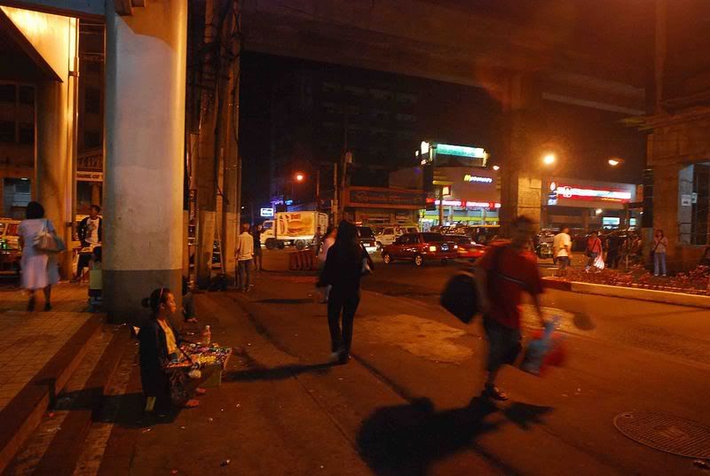
A Random Walk in Cubao @ 12 Midnight
 Cubao at midnight assumes a different personality. The darkness gives way to interesting characters in the night. I often walk in the darkened alleys and lonely side streets of Cubao, and each time is an experience of mixed fascination, disgust, and ultimately, admiration.
I belong to Cubao. No other place gives me a sense of being at home. In Cubao belongs my past, and my present. And maybe my future.
Cubao is indeed a place of utter contradictions. On one part is the new Cubao, as represented by the newly-built Gateway Mall and the new condominium buildings. But on the other part lives the old Cubao, as represented by the old familiar establishments as Ali Mall, the old Bus Terminals, the dilapidated arcades, and the side streets filled aternately with houses and bars.
Now, I take you on a tour to the Cubao that I know, the Cubao of my childhood, and the Cubao of my future.
My portraits in this series are all par below what a professional photographer could do. And maybe some images may not take your interest, such as the photo of a spilled softdrink can in a dark alley, or a scavenger dog digging the garbage, or a man pictured peeing in the public urinal.
This is what I know as Cubao, and this is what I want to share with you...and maybe through this you will have a better knowledge of what Cubao is like.
I am not a professional photographer, and my pictures lack dramatic compositions, the accepted rule of thirds, or other accepted norms of photography.
But I will let you see Cubao from the eyes of one who has been living with it....But in the end I hope you will realize that I, too, have also acquired a personality of Cubao.
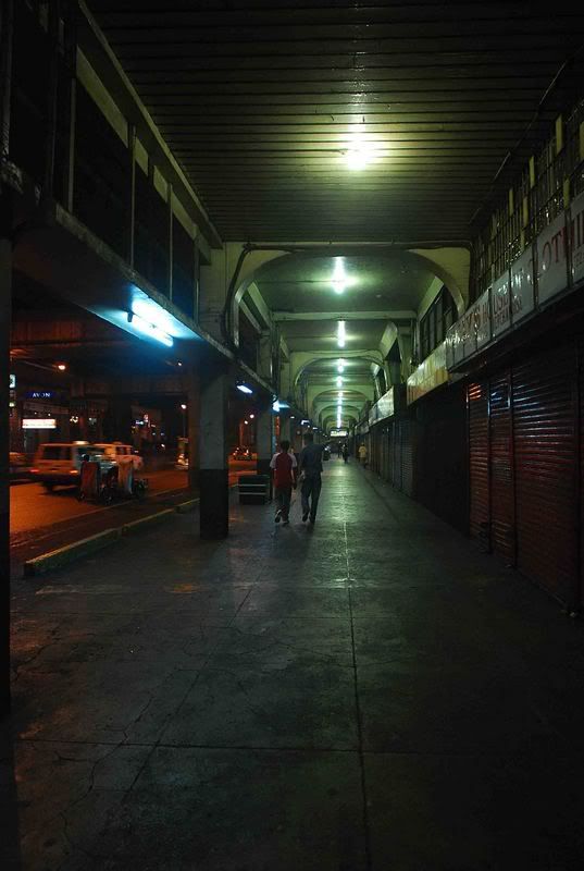
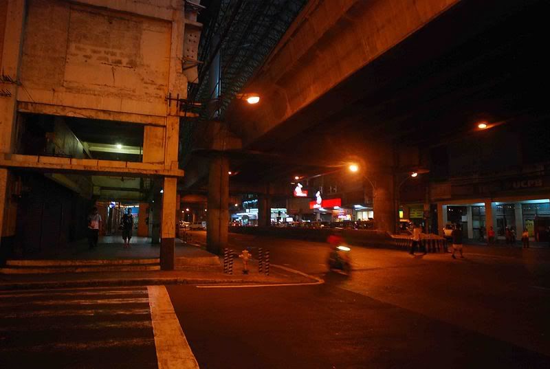 Aurora Boulevard Cubao at midnight
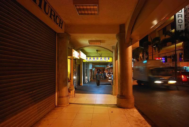 A massage spa
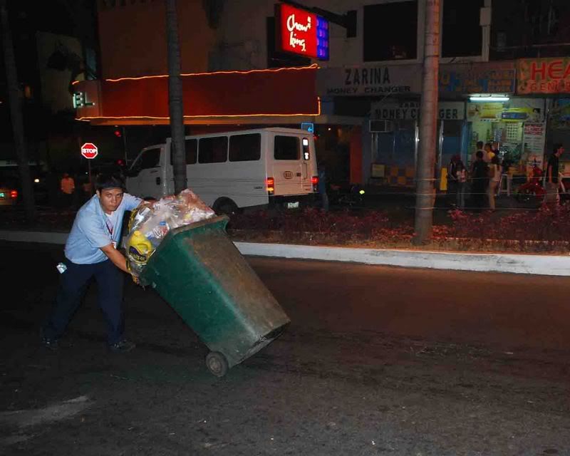 Disposing the day's garbage
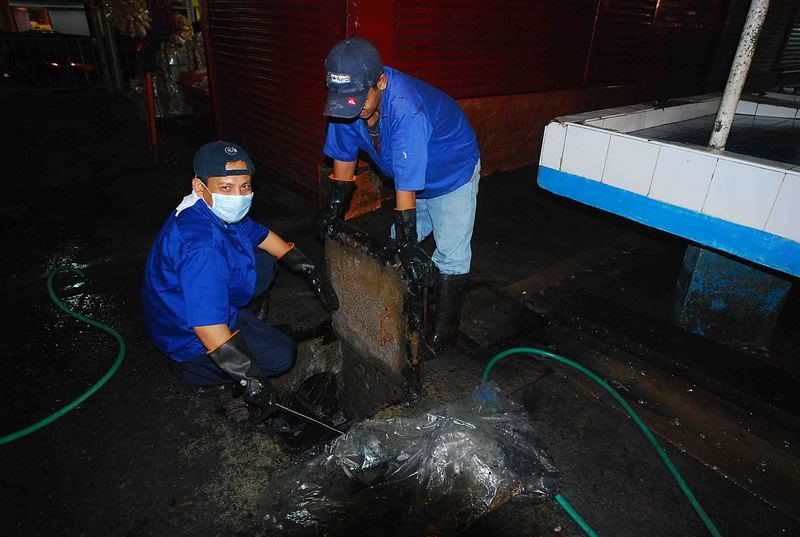 Time to clean the sewerage
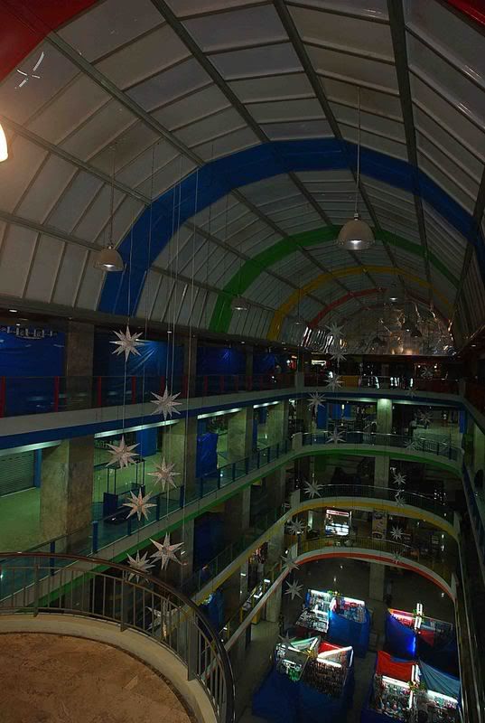 Farmer's Plaza an hour after closing
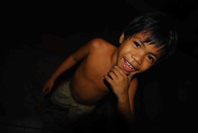 A street kid saw me photographing at night. He ran to me and asked to also be photographed. He did not ask for any money. Maybe he just felt proud to have his photo taken.
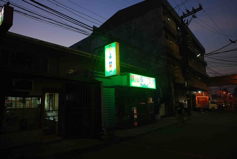 Maligamgam KTV Bar in Stanford Street
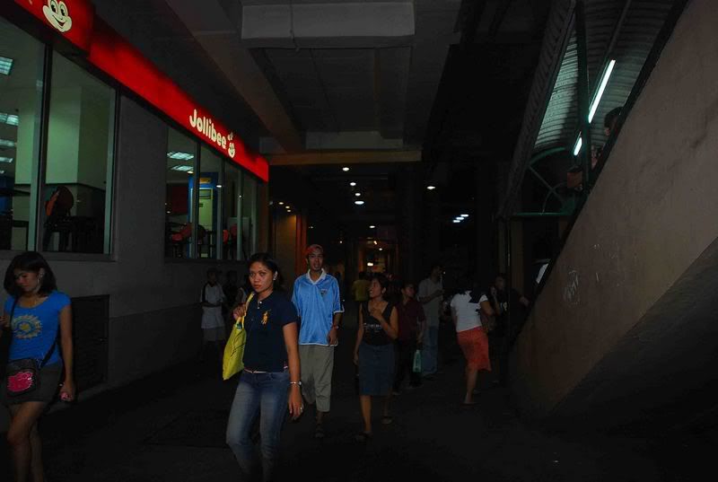
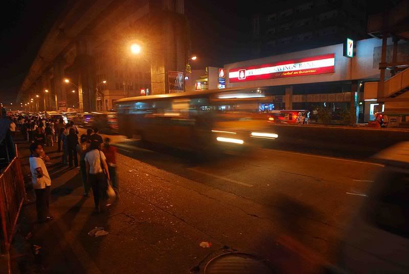 Midnight rush to go home
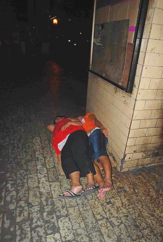 In the dark alleys of Cubao, sleep is where you can find it
 A street kid sleeping in the stair of the MRT Train station.
Trying to make a kumot out of his shirt
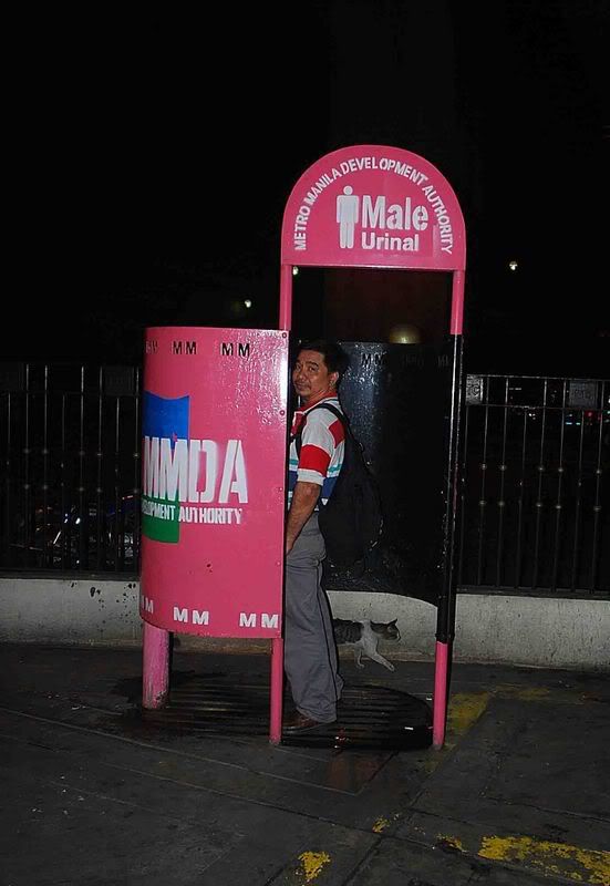 I was taking this picture of the male public urinal when this man suddenly
sensed he is being photographed. I walked away immediately.
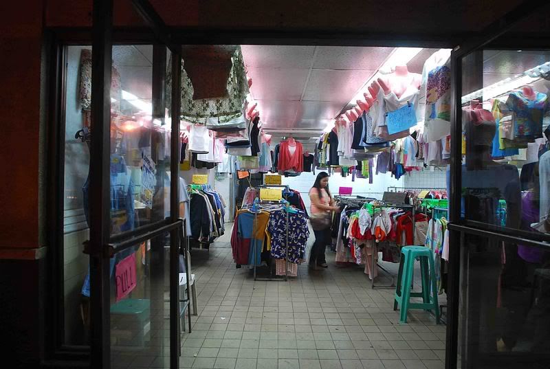 An Ukay-ukay store
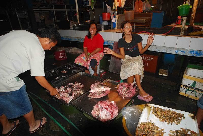 Trying to sell the unsold meat at bargain price in the
Farmer's Market, a few minutes before closing time
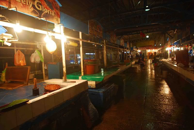 Farmer's Market closes
 An arcade in Cubao
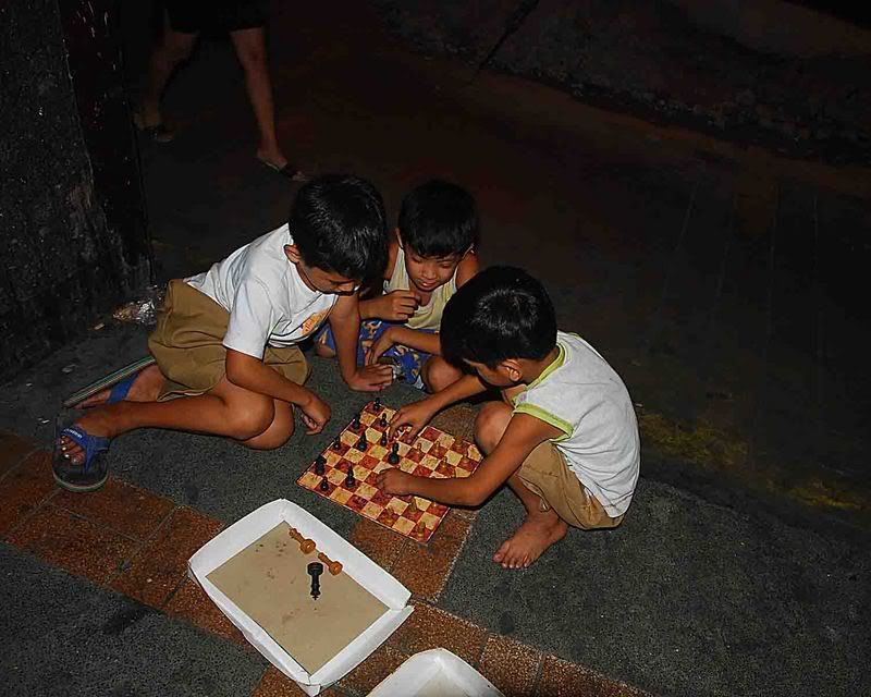 The young Chess players of Cubao. They are burning the midnight
oil to learn the strategies of the game.
 Midnight scenery
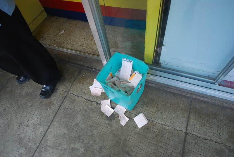 Losing Lotto tickets near the closed Lotto outlet in P.Tuazon st.
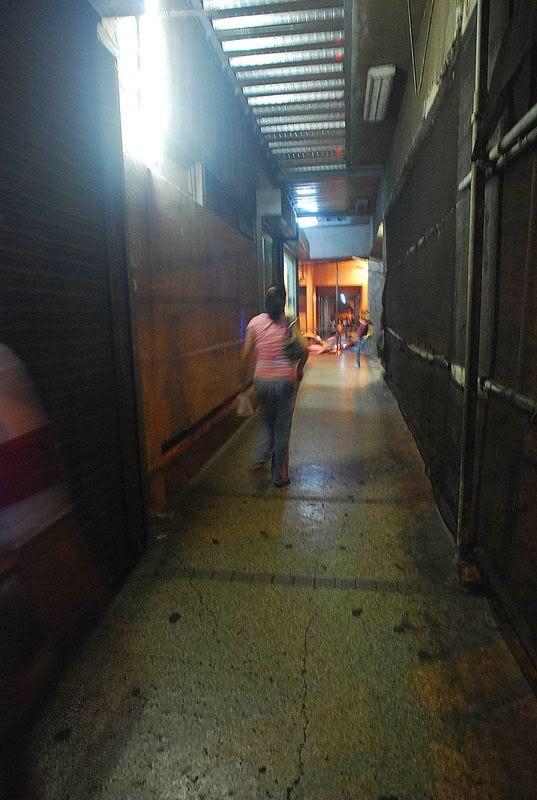 An alley in Cubao
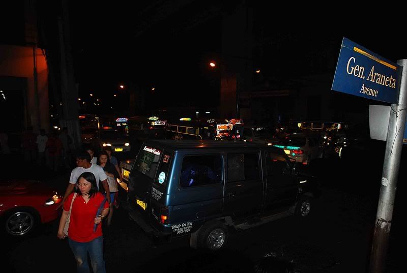 A street scene in Gen. Araneta street
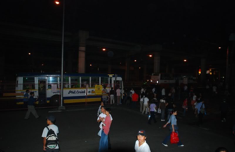 Waiting for the midnight express bus
 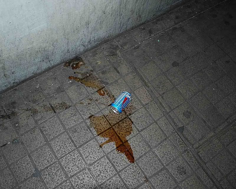 A spilled softdrink can
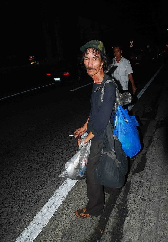 The vagrant man of Cubao
 Old cinema in Cubao, with Rated "R" movies. 35 pesos
entrance fee, and you get to see two movies. What a bargain!
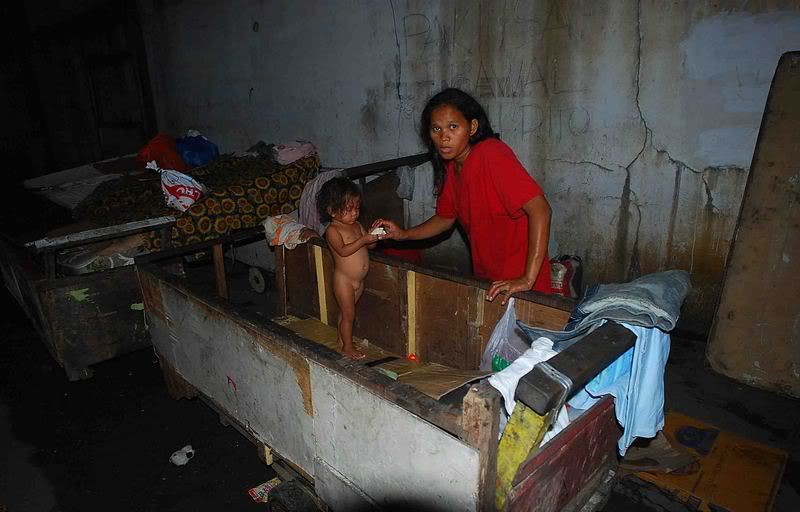 A Bahay-Kariton found behind the old COD building
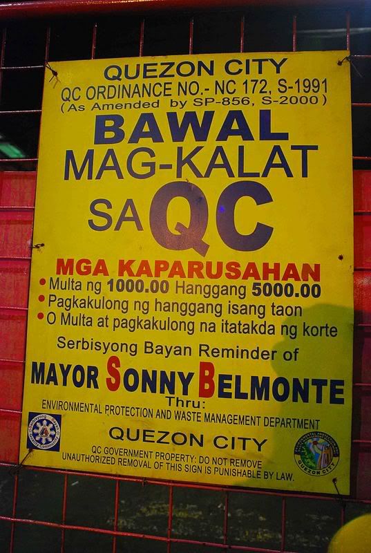 A sign to remind people
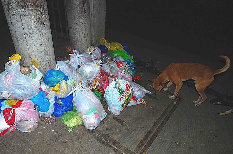 ...and the garbage.....A scavenger dog, scrapes for
left-over foods..shot near Stanford street
 Beside the famous Cubao overpass A closed arcade
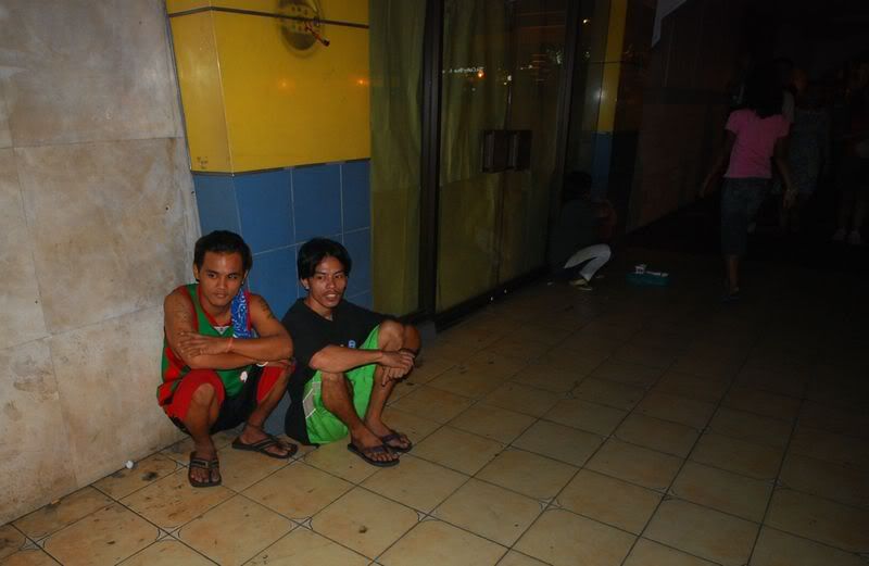 Call boys of Cubao
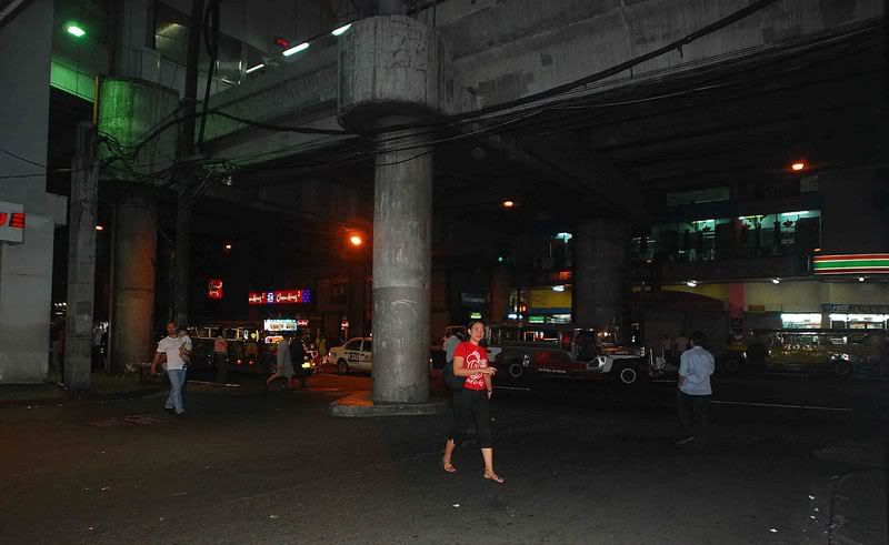 Aurora Boulevard
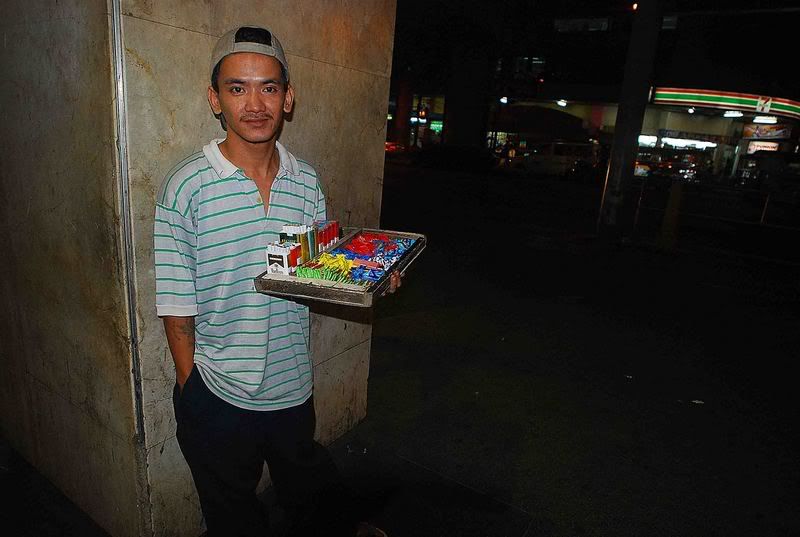 Chris sells cigarttes and candies to midnight workers and passersbys.
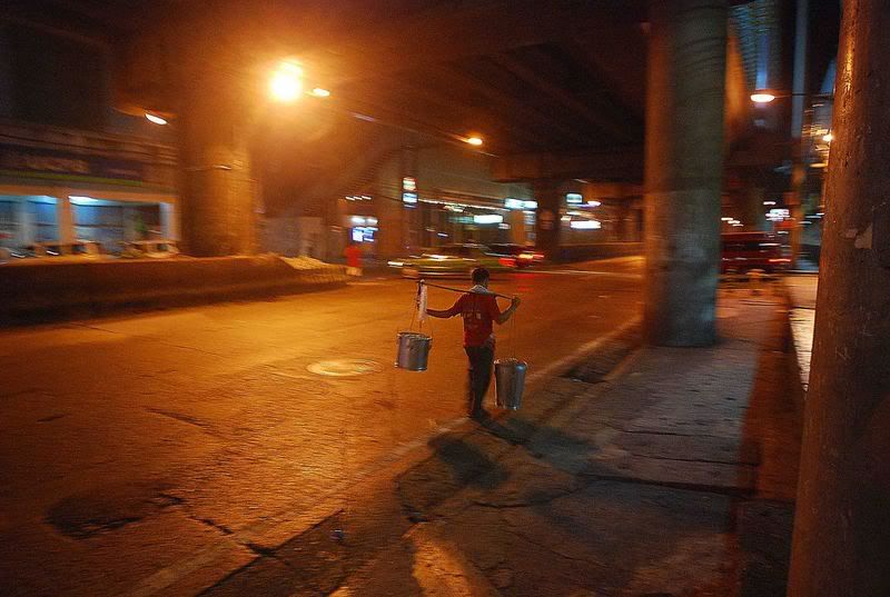 Selling Taho at midnight
Friday, January 19, 2007
Third Ventricle Arachnoid Cyst


Findings
Figure 1 and Figure 2: Axial and coronal T2 show a large cystic lesion, located within the third ventricle. There is mass effect on the surrounding normal brain structures.
Figure 3 and Figure 4: Postcontrast axial and sagittal T1 show no postcontrast enhancement of the lesion.
Figure 5: The lesion is dark (negative) on the DWI sequence.
Diagnosis: Third Ventricle Arachnoid Cyst
Arachnoid cysts form when the two layers of the arachnoid membrane split and a potential cavity is formed. The cavity fills with cerebrospinal fluid by an unknown mechanism, thought to be CSF pulsations. They can continue to grow as CSF flows in and is trapped. Arachnoid cysts rarely hemorrhage in trauma cases.
Arachnoid cysts typically cause symptoms secondary to mass effect on surrounding structures. Alternatively, they may remain asymptomatic and be discovered only as an incidental finding. Obstructive hydrocephalus, with increased intracranial pressure resulting in headaches, seizures, and developmental delay, is a common presenting feature.
Imaging findings typically feature a cystic structure with signal characteristics of CSF and dark signal on diffusion-weighted imaging; rarely, some evidence of hemorrhage may be present, especially in the setting of trauma. No solid component is present, and there is no postcontrast enhancement. There may be associated calvarial inner table scalloping, and there is typically mass effect on surrounding normal structures. Features that distinguish this entity from a subdural hygroma are convex borders, bony remodeling, and lack of substantial hemorrhage. Both entities typically cause sulcal effacement and mass effect. In cases of underlying atrophy/encephalomalacia, there would be no bone remodeling (exception is a porencephalic cyst), no surrounding normal brain architecture, and sulcal enlargement rather than effacement; cerebral veins would be seen coursing through the CSF, rather than being displaced (as with a subdural hygroma or arachnoid cyst). The lack of positive signal on the diffusion-weighted images and absence of a solid component, especially of fat signal, distinguishes an arachnoid cyst from an epidermoid, which would typically be bright on DWI.
Subscribe to:
Posts (Atom)