Friday, September 28, 2007

CNS effect of hepatic cirrhosis


Additional clinical information: Patient has hepatic cirrhosis.


Findings

There is T1 shortening of the basal ganglia, internal capsules and corticospinal tracts bilaterally. No area of abnormal contrast enhancement. There is mild cerebellar atrophy. The ventricles are normal in size and configuration. There are normal vascular flow-voids. There is no mass or abnormal fluid collection.

Differential diagnosis of T1 hyperintensity of basal ganglia:
- Cirrhosis
- TPN (manganese deposition)
- Calcification
- Products of hemoglobin breakdown (methemogbobin)
- Melanoma
- Lipid materials

Differential diagnosis (in general, abnormal intensity of basal ganglia):
- Metabolic etiologies
- Mitochondrial disorders (Leigh Syndrome)
- Methylmalonic acidemia
- Wilson's disease
- Hallervorden-Spatz
- Toxins (CO, methanol, cyanide)
- Hypoxia


Diagnosis: CNS effect of hepatic cirrhosis (Toxic-metabolic effect on basal ganglia and white matter, thought to be secondary to metal deposition)


Key points

T1 hyperintensity on MRI indicates increased fat or metallic deposition. Normally, the liver removes excess metals for storage or excretion; failure leads to hemochromatosis or Wilson's disease, in 2 examples. Another example is portosystemic shunting, as a result of liver disease (or in the case of a TIPS shunt to relieve portal hypertension). In these cases, the basal ganglia undergo metallic deposition. In the case of cirrhosis, deposition of manganese and copper lead to the bright T1 signal in the basal ganglia. In fact, degree of signal intensity quantitatively relates to the tissue concentration of manganese in the basal ganglia.

Pineal germinoma


Findings

Figure 1: Axial noncontrast CT scan demonstrates a lobulated mass of increased attenuation arising from the pineal gland. This is causing dilatation of the lateral and third ventricles.
Figure 2: Sagittal T1 demonstrates a lobulated mass arising from the pineal gland of slightly decreased signal intensity when compared to grey matter.
Figure 3: Axial FLAIR image shows mild increased signal in the region surrounding the pineal mass, representing edema.
Figure 4: Sagittal postcontrast T1 demonstrates heterogenous enhancement, which is mostly peripheral, of a pineal mass. Subtle leptomeningeal enhancement along the anterior aspect of the brainstem and the cerebral vermis is also seen.
Figure 5: Axial postcontrast T1 demonstrates heterogenous enhancement, which is mostly peripheral, of a pineal mass. Obstructive hydrocephalus is present with mild ventricular dilatation.


Diagnosis: Pineal germinoma


Pineal region tumors account for only 0.3-2.7% of intracranial tumors with germinomas making up the largest portion of the region’s tumors, approximately 40%. Additionally, germinomas are the most common of the germ cell tumors, with a large portion, 90%, presenting in patients less than 20 years of age. They tend to show a substantial male predominance. Patients with pineal germinomas may present with Parinaud syndrome (upward gaze paralysis with altered convergence). Additionally, diabetes insipidus can be a presenting sign with suprasellar germinomas prior to abnormal imaging findings.

Germinomas are more frequently located in the suprasellar region (50-60%), and less commonly located in the pineal region (30-40%). This is in contradistinction to the commonly held opinion that they occur more frequently in the pineal region. Synonyms for germinoma include: dysgerminoma, extra-gonadal seminoma, and atypical teratoma.

NECT scan of a germinoma usually reveals a mildly hyperattenuating mass that may have calcifications in or around the tumor. Both contrast enhanced CT and MRI will demonstrate intense enhancement (which is often speckled in appearance on MRI). Iso to hyperintense signal is seen on both T1 and T2 weighted MRI images. The tumor marker PLAP (Placental Alkaline Phosphatase) tends to be elevated in both the serum and the CSF of patient’s with a germinoma.

Several important diagnoses should be considered when faced with pineal region pathology. Pineal parenchymal tumors, pineoblastomas and pineocytomas, may demonstrate calcifications in or around the tumor, however this occurs three to four times more frequently with germinonas. Other lesions to consider in the differential include other germ cell tumors and tectal gliomas. Serum and CSF tumor markers, including ß-HCG and alpha-fetoprotein, may be beneficial in narrowing the differential.

Dissemination of germinomas arising from both locations into the CSF is common. Consequently, MRI of the entire neuroaxis is recommended prior to surgery. Germinomas have a relatively good prognosis because of their sensitivity to radiation and chemotherapy; 5-year survival approaches 90%.

Thursday, September 27, 2007

Hallervorden Spatz Disease-A Rare Case Report


This patient was referred to us for second opinion with clinical features of dystonia and non-specific initial MRI report. On MRI we noted bilaterally symmetric hyperintense signal changes in anterior medial globus pallidus with surrounding hypointensity in the globus pallidus on T2-weighted images. These imaging features are fairly diagnostic of HSD and have been termed the “eye-of-the-tiger” sign.The hyperintensity represents pathologic changes including gliosis, demyelination, neuronal loss, and axonal swelling, and the surrounding hypointensity is due to loss of signal secondary to iron deposition.


Monday, September 24, 2007

Sumer's Radiology Site features in SERAM, Sociedad Española de Radiología Médica

SERAM
http://www.seram.es/
Sociedad Española de Radiología Médica


Sumer's Radiology Site figures in the websites of interest column.


Glioblastoma multiforme


Findings

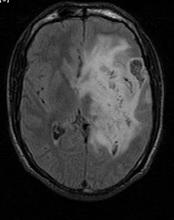
CT shows a large region of primarily white matter mass-like with hypodense center, with mild mass-effect in the high left frontoparietal region. MRI confirms a mass within the left frontoparietal region. This mass demonstrates thick, irregular peripheral post contrast enhancement and surrounding edema. There is no restricted diffusion within the central region of necrosis. There is no decreased T2 rim signal to suggest a fibrotic capsule. There is mild mass-effect on the left lateral ventricle and minimal left-to-right midline shift.


Diagnosis: Glioblastoma multiforme (biopsy proven)


Each year approximately 17,000 primary brain tumors are diagnosed in the United States. Sixty percent of these tumors are gliomas. Glioblastoma multiforme (GBM) is the most common and most malignant of the gliomas. The GBM is a heterogeneous tumor composed of poorly differentiated astrocytes. Most commonly, GBMs affects adults and involve the cerebral hemispheres. Although much less likely, GBMs can affect the brain stem and spinal cord in children. GBMs can transform from lower-grade astrocytomas or anaplastic astrocytomas. However, in most instances, GBM arises new, without a precursor lesion. The GBM can spread via several routes including: white matter tracts, across the midline via commissures such as the corpus callosum, subependymal seeding of the ventricles, and CSF seeding of the subarachnoid space.

In this case, the differential diagnosis includes both neoplasm (most likely glioblastoma multiforme) or abscess. Both diagnoses were considered almost equally after the initial head CT. The MRI provided some new information, but still was not be conclusive for either neoplasm or abscess. Specifically, the lesion demonstrated a central region of necrosis but lacked restricted diffusion, making abscess less likely. Similarly, the lesion lacked a decreased T2 rim signal to suggest a fibrotic capsule, which also made abscess a less likely diagnosis. Because imaging was not conclusive for neoplasm or abscess, stereotactic biopsy was performed and revealed glioblastoma multiforme.


Radiologic overview of the diagnosis

Usually heterogeneous low-density mass (seen on CT)
Intense contrast enhancement
Hemorrhage and central necrosis are common
Calcification is uncommon
Abundant vasogenic edema
Mass effect
Bihemispheric spread via the corpus callosum or commissures (the classic "Butterfly lesion")
If peripheral location – often demonstrate broad dural base and dural tail (appearance similar to an extra-axial lesion)
CSF seeding (leptomeningeal drop metastases)

Sunday, September 23, 2007

Radiology Quiz-What is the Diagnosis?


This patient presented with complaints of vomitting and abdominal pain. Can you make the diagnosis on the basis of scout film and CT section.


Post your answers in the comments section. Case reported by emergency team of Teleradiology Providers

Answer--
Intussusception


Winner-
Dr. Eytan RAZ

The Koreans in the Philippines

Yesterday, while dozing off to a boring comedy movie in Ali Mall, I was suddenly awakened by a loud burst of laughter coming from a group of viewers behind my chair. I found out that these laughing people were not Pinoys at all, but Koreans. They were really enjoying the movie. It was curious though that they were the only ones laughing, while the rest of the viewers maintained their silence.

I have long noticed the swelling numbers of Korean tourists in our country. They are all over the Philippines, in the parks, beaches, resorts, hotels, and everywhere. Now, the Koreans are starting to invade the malls.

If you had been to Luneta or Intramuros lately, you would notice several identical tourist buses parked in the side streets, all rented by Koreans. Most of them were all in walking shorts, white shirts and caps, and everyone carries a digicam. They take pictures of everything, as in everything. In my observation, they do not mingle with the locals, only limiting their contacts with their Filipino tourist guide.

One late afternoon sometime in January this year, I decided to go to Manila Bay breakwater, where I can ride in one of those for-rent passenger boats for 50 pesos, and experience Manila Bay sunset at the sea. I climbed inside the boat and waited several minutes for other passengers to fill in the vacant seats. Three or more Pinoy couples entered the boat. Suddenly, we were informed by the boat owner that we had to vacate the boat as it had suddenly been rented "wholesale" by Korean tourists. I complained that I can also pay the 50 pesos fare, and that I didn't mind riding with the Koreans. The boat owner told me that the boat would be "exclusively" for Koreans only. Okay, so the Koreans didn't want Pinoys aboard. Alright then. Let them have the boat. I can just enjoy my sunset sitting in one of the benches.

Another incident that involved me and Korean tourists happened last July, 2007. I was in Intramuros to buy some books at the Ilustrado Bookshop. Upon entering the shop, the Filipino guard at the door asked some me to inspect my bag. I thought it was s.o.p. so I showed him the contents of my bag, no problem. After the guard thoroughly inspected my bag, three Korean tourists (I tell you, they're everywhere) entered the shop. The guard immediately greeted them in a warm manner (not accorded to me), and let them inside the shop without inspecting their huge bags. I was furious at this discrimination and reprimanded the guard for his toady behavior in front of foreigners. He got my point and apologized. The Koreans looked bewildered.

As Filipinos, we should stand on equal footing with foreigners, especially in our own country. Subservient behavior in front of foreigners does not help in the prestige of our image as Filipinos. Otherwise, foreigners would think we are only after their money. It is good to be hospitable but we should always remember to maintain dignity and self-respect.

The Growing Population of Koreans in the Philippines

According to the latest stats, the number of Korean tourists in the Philippines ballooned from 378,602 in 2003, to 572,133 in 2006, a 51-percent rise.(Source: "Koreans "invade" the Philippines, Philippine Daily Inquirer 6/17/2007)

The figures may have certainly grown by now, as the Philippine government allows, even encourages, foreigners to come to our country. Their visits earn huge amounts of income for the government. But what will the effect be in the long run?

There is a variety of reasons why Koreans favored the Philippines over other Asian destinations. The Philippines' lax in immigration requirements, the low cost of travel and expenses in the Philippines, hospital tourism, and the low cost of education.

Many of the Koreans who were granted alien certificate of residence in the Philippines already acquired vast amount of real properties. Everywhere in the country, they have already made their presence felt, establishing exclusive schools for Koreans, resorts, restaurants, hotels, and even hospitals for Koreans.

What's surprising is that foreigners are not supposed to own real properties in the Philippines. If they do, they are only entitled to be a minor owner only--meaning there should be a major Filipino co-owner.

Recently, some concerned Filipinos exposed that a wealthy Korean company bought a large tract of land in Taal, Batangas. The land was planned to be used as a resort spa. It was found that the Mayor of Talisay issued the memorandum of agreement for the purchase, even though it is clear that resort spas within the outskirts of the Taal volcano are not allowed to be built.(source:William Prilles Jr."The Furor on the Taal Spa" @http//www.planet.naga.gov)

In Baguio City, residents were alarmed at the growing rate of Korean residents in the city. According to residents, the common greeting nowadays is "Anyung Ha Seyo"--Good morning in Korean. Angela Malicdem of Bulatlat.com reports that, "In Baguio, the influx of Korean nationals has caught some attention. Baguio now is host to almost 10,000 Koreans. At first, only teenagers came here, to study the English language. Most of them stayed for two months during their vacation from school in Korea. Then they started to study full-time in Baguio universities. Before long the Koreans started coming to Baguio with their whole families. "

Now, I don't have anything against Korean tourists, the Korean people or the Korean nation. I am no xenophobic, and in fact I admire the Koreans and how they have managed to deal with the difficult separation of their country and still maintain their unique culture. However, it causes me some alarm that now, there is definitely an ongoing silent invasion of Koreans in the Philippines. And many of these Koreans are not tourists anymore. Many have already started to settle permanently.
Saturday, September 22, 2007

Radiology Grand Rounds-XVI


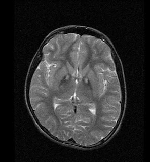
Here is a case of Pineal tumour for the Radiology Grand Rounds submitted by Dr MGK Murthy, Dr Sumer Sethi of Teleradiology Providers. Concept of the Radiology Grand Rounds is available at- Radiology Grand Rounds.


The approach to such cases--
Germinoma and teratoma are two germ cell tumors that can arise from the pineal gland. Germinoma is the most common pineal tumor, accounting for 40-50% of all pineal region tumors and two-thirds of all germ cell tumors. It affects primarily children or young adults and is significantly more common in males. Germinomas have a homogenous appearance on MRI and enhance strongly following contrast administration. Teratoma accounts for 15% of pineal masses and also demonstrates male predominance. These lesions are heterogeneous with calcifications and mixed CSF, lipid and soft tissue areas. Germ cell tumors tend to engulf primary pineal calcifications.
Pineal parenchymal lesions include pineocytoma and pineobalstoma. Pineocytoma is an enhancing lesion that affects adults. This benign entity is characterized by good demarcation, homogeneity, and slow growth. It is noninvasive. Pineoblastoma generally affects children and occurs with approximately equal incidence in males and females. Common characteristics of this malignancy include local invasion, distant CNS spread, heterogeneity, calcification, and marked enhancement. It is important to obtain images of the spine to look for metastases. Parenchymal tumors will have intrinsic calcifications, producing an exploded appearance .
The above case is a lady young and homogenous enhancement hence pineocytoma is possible.
The clinical presentation of a pineal lesion may include obstructive hydrocephalus, Parinaud's syndrome (characterized by palsy of upward gaze, dissociation of light and accommodation, and failure of convergence) caused by compression of the tectum, and endocrine abnormalities ( e.g., precocious puberty) in cases of germ cell tumors. One of the radiologist's main roles is to determine the origin of the lesion. Lesions arising from adjacent regions and extending to the pineal region include CNS lipoma, epidermoid, arachnoid cyst, astrocytoma, and meningioma. Lesions arising from the pineal gland itself include germ cell tumors, pineal parenchymal lesions, pineal cysts, vascular malformations, and metastases.

Tuesday, September 18, 2007

Dyaran! My Dinner Tonight!

I came home starved after driving for an hour in traffic congested EDSA. I searched for food in the fridge but found mostly fresh meat and jugs of water.
And so, I just bought a 12-peso Sardines from our local Sari-sari store. I never liked Sardines at all. But wouldn't you know it, it tasted really good together with frozen rice...Well, everything should be delicious to a hungry stomach.

Supraclinoid Internal Carotid Artery Aneurysm--MRI


Here are images of a case of supraclinoid ICA aneurysm on MRI sent by Dr MK Murthy, Dr Sumer Sethi, Dr David, Dr Padamaja Teleradiology Providers

Cryptococcal meningitis


Findings

There is nodular thickening and T2-weighted hyperintensity of the callosal splenium with associated diffusion restriction. There is subtle leptomeningeal enhancement.

Differential Diagnosis:
- Embolic infarcts
- Abscess / multifocal infection
- Marchiafava-Bignami syndrome
- Diffuse axonal injury
- Lymphoma


Diagnosis: Cryptococcal meningitis


Key points

Cryptococcus neoformans is a ubiquitous yeast that grows abundantly in soil containing bird (especially pigeon) droppings. Disseminated infection occurs mainly in the immunocompromised, with the lungs and central nervous system most commonly affected. Cryptococcosis risk factors include; AIDS, steroid therapy, immunosuppression induced after organ transplantation, lymphoproliferative disorders, diabetes, tuberculosis, and sarcoidosis. Approximately 30-50% of patients with cryptococcal meningitis, however, show no evidence of immunosuppression (as in this case).

Nonspecific symptoms include:
- Fever
- Headache
- Changes in level of consciousness (somnolence, confusion, stupor or coma)
- Dizziness
- Visual disturbances
- Seizures

Diagnosis relies on CSF Indian ink preparation, culture, and antigen titers.


Radiologic overview of the diagnosis

Manifests as meningoencephalitis with gyral enhancement and intraparechymal lesions. Intraparechymal lesions include non-enhancing gelatinous pseudocysts (often in the basal ganglia) and enhancing cryptococcomas. Cerebral infarction can result from chronic infection. Brain lesions may worsen with Amphoterician-B treatment, causing concern for treatment failure. Hydrocephalus and diffuse atrophy can also be seen, yet could be related to an underlying disease process such a HIV.

Nasopharyngeal carcinoma


Findings

Figure 1: Axial CT image displayed at bone window reveals the destructive lesion at the skull base involving the basi-sphenoid, basi-occiput, pterygoid plates and extending across the midline.
Figure 2: Axial T2-WI reveals soft tissue mass in the right lateral pharyngeal recess of the nasopharynx, involving the right veli palatine muscle and effacement of the Fossa of Rosenmüller.
Figure 3: Axial T2 WI reveals the mass to extend superiorly involving the right pterygopalatine fossa and the right pterygoid recess. The right masticator space appears spared.
Figure 4: Axial T2 WI reveals the mass to extend to involve the right sphenoid sinus and petrous apex, and into the right cavernous sinus, encasing and narrowing the right cavernous carotid artery.
Figure 5: Axial contrast-enhanced T1-weighted image obtained at level of nasopharynx shows fullness and enhancement in region of right Rosenmüller's fossa.
Figure 6 and Figure 7: Axial contrast-enhanced T1-weighted images reveal extension into the sphenoid sinus, petrous apex, pterygopalatine fossa and right cavernous sinus.
Figure 8: Coronal contrast-enhanced T1-weighted MR image reveals invasion of nasopharyngeal cancer into right cavernous sinus with encasement of internal carotid artery.
Figure 9: Coronal contrast enhanced T1-weighted image reveals an enhancing mass in sphenoid sinus region and increased enhancement (compared with normal left side) extending through the right foramen ovale.


Diagnosis: Nasopharyngeal carcinoma


Most nasopharyngeal malignancies in adults are carcinomas, usually of the squamous cell variety (80%). Squamous cell carcinomas (SCC) of the nasopharynx are relatively rare in the United States and much more common in Asia, being endemic in Southern China. A strong correlation between Epstein-Barr virus and nasopharyngeal cancer is well documented. Genetic predisposition and association with consumption of salted fish and preserved foods is also suspected.

Nasopharyngeal squamous cell cancer usually presents before age 50 and is more common in males (M:F=2.5:1). It usually presents late because of its infiltrative pattern. The most common presentation is an asymptomatic neck mass due to metastatic adenopathy. Serous otitis media may result from eustachian tube dysfunction resulting in otalgia and unilateral conductive hearing loss. Nasal obstruction and epistaxis are other common symptoms. Perineural spread and intracranial extension can cause headaches and cranial nerve deficits.

Nasopharyngeal cancers usually present as a poorly marginated nasopharyngeal mucosal space mass with deep extension and invasion. They tend to grow along the path of least resistance: along submucosal and soft tissue planes, along neurovascular bundles and intracranially through neural foramina at the skull base. Nodal metastases are present in 90% of cases at presentation. Distant metastases to the bones, lung or liver are seen in less than 10% of cases.

These tumors show squamous differentiation with intracellular bridges or keratinization. There are three main histologic subtypes- keratinizing, non-keratinizing and undifferentiated carcinoma.

Biopsy is the way to establish the diagnosis since imaging cannot distinguish among nasopharyngeal malignancies. Imaging plays a crucial role for mapping out the spread of tumor and planning radiation therapy/ surgical resection. Enhanced MR imaging is the best tool for evaluating intracranial extension via direct, perineural or perivascular routes. Thin section CT helps to evaluate bone invasion. CECT is preferred for evaluation of cervical metastatic nodes, except retropharyngeal adenopathy which is better seen on MRI. These tumors are strongly FDG avid.

XRT is the mainstay of treatment with combined XRT and chemotherapy used for advanced disease.

Monday, September 17, 2007

Photographing the Photographer

I took this photo exactly a year ago during the Hidalgo Photographers Haven Contest sponsored by the City of Manila. I submitted this as my entry in the category: "Photographing the photographer". It did not win, of course, but at least I made some of the judges smile.. hehe :)

Friday, September 14, 2007

Searching for Caparas' Komiks

I left office early afternoon yesterday to go to Quiapo to hear the traditional Friday mass. Anyway, the second reason for going there is to buy the new komiks titles of Carlo J. Caparas. It had been months that I'm anxiously awaiting these new komiks and I can't get them off my mind even while trying to hear Father Ramirez' homily.

After the mass, I went to Ma Mon Luk in Quezon Blvd. to eat as I'm already feeling hungry. On the way, I kept on looking at the newsstands to buy Carlo Caparas' Komiks. The sellers told me that they were sold out. And so I arrived in Ma Mon Luk still without my komiks.

My waiter is already waiting for me as he knew that I usually eat there after hearing the evening mass.

I ordered Special Siopao (50 pesos) and regular Mami (80 pesos) and Coke (15 pesos). The food is delicious, but I can't help but feel that I was dining in one of the old cafes in Manila during the 1950s.
The old ambience, with the antique ceiling fans and old pictures hanging in the walls give Ma Mon Luk restaurant an antique look.

After dining, I went to search once more for Carlo Caparas' Komiks. As it turned out, most of the newsstands have sold it out a few hours ago. Apparently some guy bought all their copies with the intent of hoarding them. At 10 pesos each copy( and the five Caparas komiks are all Vol.1 No.1.), I understood the reason.

But I can't just give up. There should still be some copies left here in Quiapo, and I'll turn downtown Manila upside down to find them. I need to find them to update my microsite in Wikipilipinas. I'm currently building a komiks encyclopedia and almanac within Wikipilipinas. Now I won't let another person become the first to put on the Wikipilipinas information on these Caparas' Komiks. If there is one person to show them in Wikipilipinas, it should be me.

Fortunately, a nice seller told me to go to Palanca because he thought he still saw some copies there. I went immediately otherwise they may be bought by someone else.

I found the seller, but alas, no more copies left. He told me though that he still have some copies he was reserving to someone else. If I want to buy them, then he'd sell them to me for 20 pesos per copy. Deal or no deal?

Now, I don't usually deal with greedy people, but as it was already getting late, and I need to update my Wiki komiks encyclopedia, I agreed even if paying double. And now, I finally have the complete 5 komiks titles of Caparas. I already uploaded their preview in Wikipilipinas.

Here they are:

Gwapo Komiks Vol.1 No.1


OFW Super Stories Vol.1 No.1


Super Funny Komix Vol.1 No.1


Klasik Komiks Vol.1 No.1

If you want to know more about these komiks, buy them (if copies are still available), or visit Wikipilipinas for a preview.

Thursday, September 13, 2007

Infected second branchial cleft cyst


Findings

There is a well circumscribed low attenuating, likely cystic, mass located posterior to the submandibular gland, lateral to the carotid space, and anteromedial to the sternocleidomastoid muscle. The mass demonstrates slight rim enhancement, with mild inflammation of the adjacent fat.

Differential diagnosis:
- Infected second branchial cleft cyst
- Necrotic malignant adenopathy
- Lymphangioma
- Thymic cyst


Diagnosis: Infected second branchial cleft cyst


Key points

Most common type of branchial cleft anomaly – accounts for 75% of branchial cleft anomaly in children and >90% in adults
Results from failure of obliteration of the cervical sinus
Usually not associated with other anomalies
Four subtypes depending on location: Bailey classification – type II is the "classic" location as described below
May have associated fistulas

Clinical Presentation:
- Presents as painless mass at the lateral aspect of the neck
- Mass may be chronic, and may become larger with respiratory infection
- May be painful if infected
- Age of presentation between 10 and 40 year old
- No sex or race predilection
- Treatment is complete surgical excision, with good prognosis
- Controversial if malignancy can arises from branchial remnant


Imaging

Usually a round or oval shaped cystic mass
Located at the anterior triangle or angle or mandible
Lateral to carotid space, posterior to the submandibular gland, and anteromedial to the sternocleidomastoid muscle
Variable in size, may be up to 10 cm

Contrast enhanced CT:
- Cystic density mass with non enhancing wall
- If infected, wall will be thicker and may enhance, with inflammation of adjacent soft tissues

MRI:
- Cystic mass isointense to fluid on T1 and T2
- May become hyperintense on T1 if infected
- Will demonstrate wall enhancement if infected

Wednesday, September 12, 2007

Gliomatosis Cerebri-MRI & MRS


Gliomatosis cerebri (infiltrative diffuse astrocytosis) is a rare primary brain tumor. It is commonly characterized by diffuse infiltration of the brain with neoplastic glial cells that affect various areas of the cerebral lobes. Before the advent of MRI, diagnosis was generally not established until autopsy. Typically, gliomatosis cerebri appears as a diffuse, poorly circumscribed, infiltrating non-enhancing lesion that is hyperintense on T2-weighted images and expands the cerebral white matter. The prognosis for gliomatosis cerebri is generally poor, with a median survival time of only 12 months.

Mucopolysaccharidoses (MPS) / Hurler disease


Findings

Figure 1: T1-weighted image showing dilated perivascular spaces.
Figure 2: Abnormal T2-signal in surrounding white matter. T2-weighted image showing dilated perivascular spaces.


Diagnosis: Mucopolysaccharidoses (MPS) / Hurler disease


Mucopolysaccharidoses (MPS) are inherited disorders of metabolism which are characterized by the inability to break down glycosaminoglycan (GAG) because of an enzyme deficiency. This results in an accumulation of toxic intracellular substrate. There are multiple MPS, classified from MPS 1-9, depending upon the specific enzyme deficiency. The prototype is MPS 1H, which is Hurler disease. This is caused by alpha-L-Iduronidase deficiency.

On noncontrast CT there is macrocrania. The dilated perivascular spaces are rarely visible on CT. There is progressive hydrocephalus and atrophy. On contrast enhanced CT there is often enhancing pannus associated with the ligaments and dura at the craniocervical junction.

A classic diagnostic imaging finding on MRI is dilated perivascular spaces (PVS) or Virchow-Robin spaces. This will appear as dark on T1WI and bright on T2WI. The dilated PVS are usually under 5 mm, but can be larger, and range from one to many. The most common locations are the corpus callosum and peritrigonal white matter, but can occur anywhere. On T2WI, there is often increased signal of the white matter surrounding the dilated PVS, which is a result of gliosis, edema, demyelination or dysmyelination. On FLAIR, there may be increased signal surrounding the PVS.